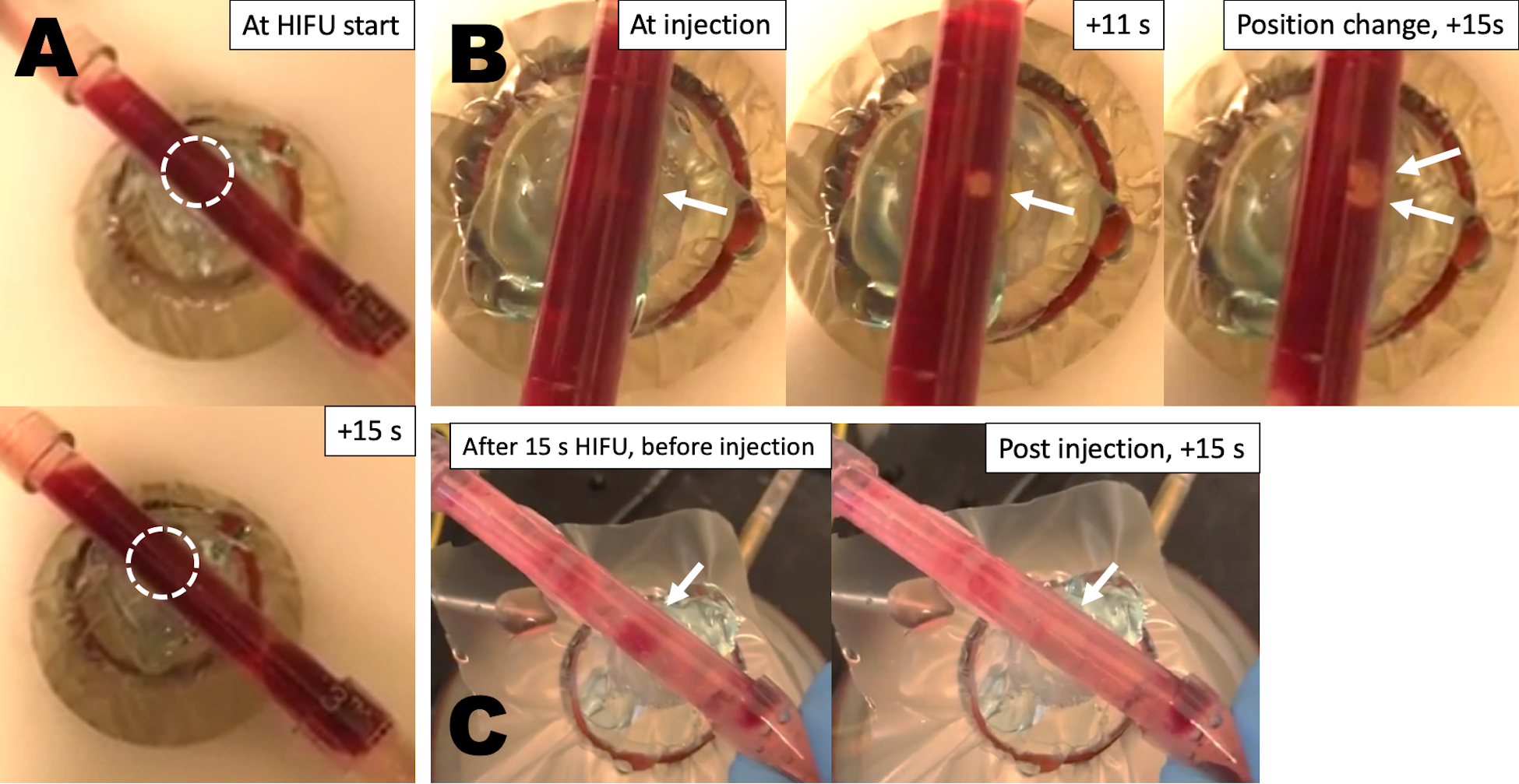
Figure 5.

Still images from videos of the in vitro thrombolysis flow model. (A) Image of partially occlusive thrombus before and after HIFU insonation without injection of particles. (B) Images of the same blood clot at administration of P@hMSNs, then after 11 s of HIFU insonation, and once more after a position change and an additional 15 s of HIFU. (C) Another example of blood clot after 15 s of HIFU prior to P@hMSN injection, then again post injection with 15 s HIFU.
