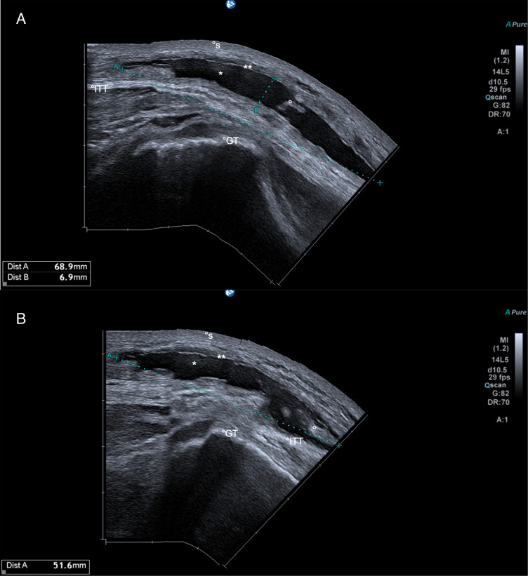
Figure 1.
Utrasound scan performed using a linear transducer (12 MHz) showing long-axis supratrochanteric view (A), and short-axis supratrochanteric view (B). The images demonstrate a homogenous anechoic fluid collection (*), measuring approximately 7×0.7 x 5 cm in diameter, associated with scattered hyperechoic substance (°) between subcutaneous tissue (**) and the Ilio-tibial tract (ITT). Annotated additional structures: GT, greater trochanter; S, skin.

