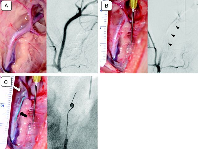
Fig 2.
A, Photograph of a surgically exposed left superficial cervical artery (left) and its selective angiogram (right). Note that multiple branches of the superficial cervical artery are seen in both the surgical view and the angiogram. B, Intraprocedural photograph (left) and an angiogram (right) of the left superficial cervical artery (postocclusion). The left superficial cervical artery shown in A was occluded with an injected experimental thrombus (left). Pulsation of the artery diminishes significantly after the occlusion, and the artery becomes pale. An angiogram was performed. C, Intraprocedural photograph (left) and an angiogram (right) of the left superficial cervical artery (Merci device deployment). A Merci clot retriever, deployed at the postbifurcation segment of the left superficial cervical artery (open arrow) and a microcatheter (arrow) are seen through the wall of the vessel (left). A fluoroscopic view of the same artery shows the device being deployed and reformed into its original coil shape (right). Behavior of the deployed device can be monitored during the procedure under direct vision.

