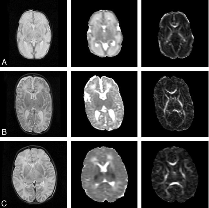
Fig 1.
Anatomic, MD, and FA map of a neonate. A, At 30 weeks' gestational age; B, at term age; and C, after perinatal asphyxia scanned on day 4, with abnormal low signal intensity in the central gray matter on the MD map. Notice the decrease in MD values and increase in FA values between the preterm and a term brain. The MD maps and FA maps are equally scaled for the 3 subjects, respectively, 0–2 × 10⁻³ mm2/s and 0–1.

