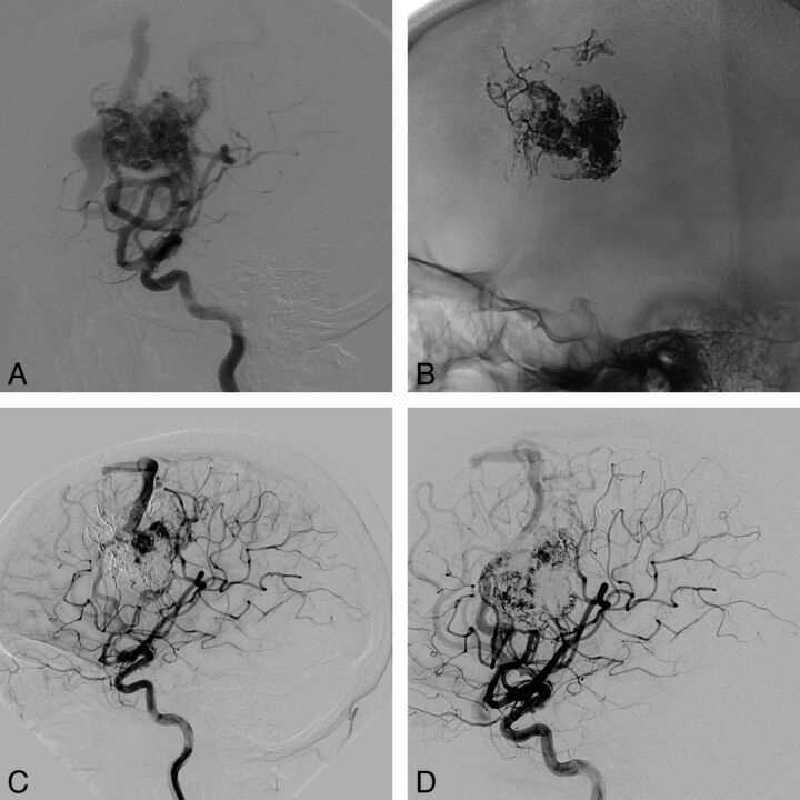
Fig 2.
Right frontal AVM. A, Right internal carotid angiogram (lateral view) shows the AVM before treatment. B, Right internal carotid angiogram, noninjected and unsubstracted (lateral view), shows the Onyx cast after 5 embolization sessions and before radiosurgery. C, Right internal carotid angiogram shows the residual nidus before radiosurgery. D, Right internal carotid angiogram shows a residual nidus 4 years after radiosurgery.

