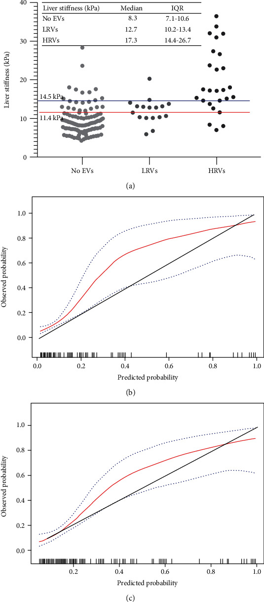
Figure 2.

Distribution of liver stiffness was subdivided based on the absence of EVs, presence of LRVs or HRVs in all patients (a). The red and blue horizontal lines represent cut-offs of 2D-SWE for varices of all sizes and high-risk varices, respectively. Calibration plot of 2D-SWE (bootstrap resampling times = 500) for high-risk varices and varices of all sizes in all patients (b) and (c). Calibration slopes graph the agreement between predicted probability on the x-axis and observed proportion on the y-axis. The black dashed line represents perfect calibration, with 100% agreement. Red line represents the performance of 2D-SWE, and blue dashed line represents 95% confidence intervals of observed probabilities. Abbreviations: EVs: esophageal varices; LRVs: low-risk varices; HRVs: high-risk varices; 2D-SWE: two-dimensional shear wave elastography; kPa: kilopascals.
