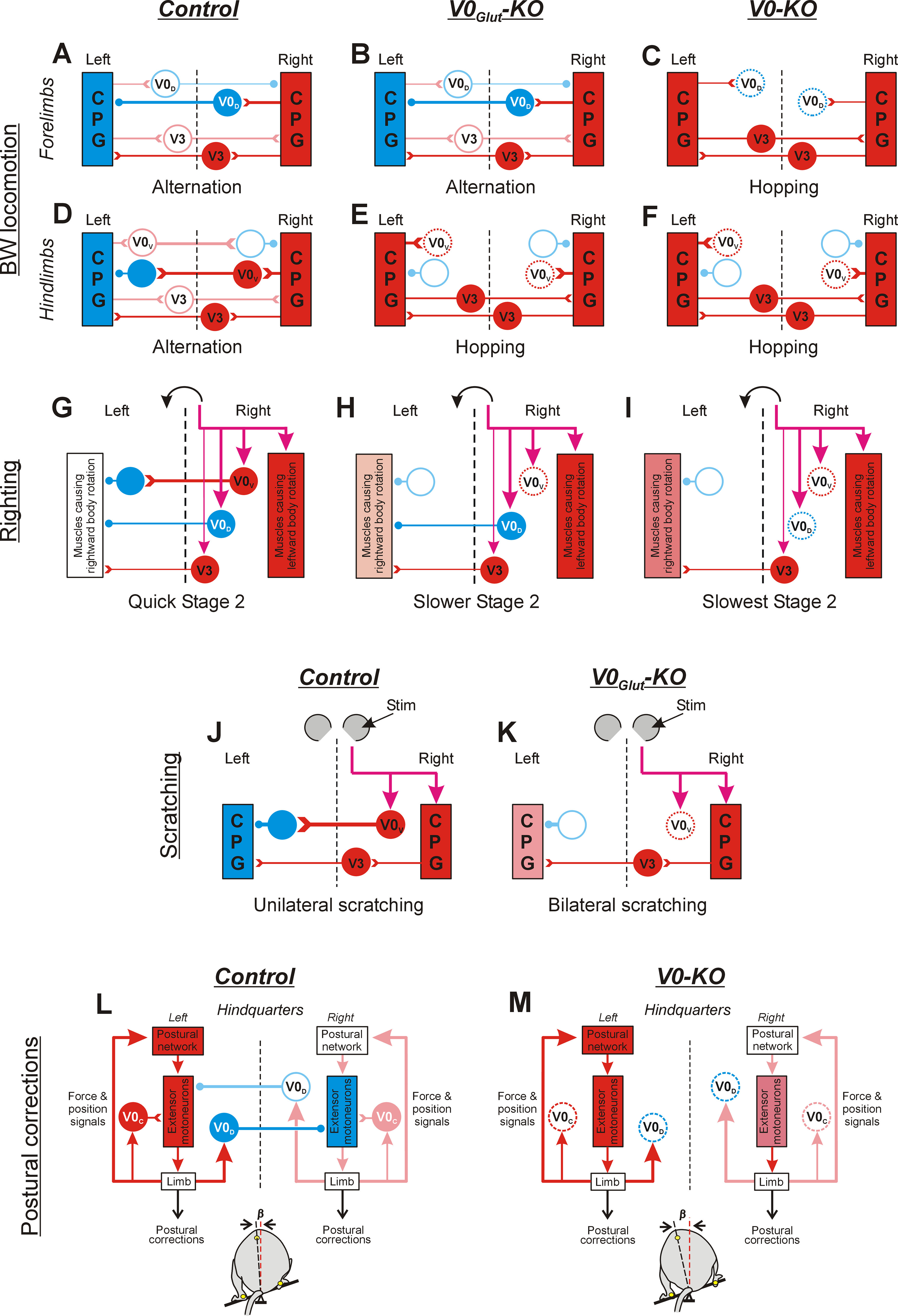
Figure 5.

Diagrams depicting the changes in network configurations as inferred from the ablation of V0V and all V0 CINs. Designations are as follows: activated inhibitory and excitatory neurons are shown in blue and red, respectively; inactive inhibitory and excitatory neurons are indicated by light blue and pink empty circles, respectively; ablated neurons are indicated by dotted circles; progressive increase in excitation level is indicated by a gradual increase in the intensity of red color. Open triangles, excitatory synapses; small filled circles, inhibitory synapses. The thickness of lines indicates the strength of the produced effect. Only CINs contributing to the generation of a behavior (as inferred from the perturbed behavior in KO mice) are shown. In A–F, J, and K, for simplicity, only parts of left and right CPGs generating the same phase of the cycle are shown. Purple arrows indicate descending commands causing the behavior. A–F, Proposed networks underlying the coordination of forelimbs (A–C) and hindlimbs (D–F) during BW locomotion, in control mice (Control; A, D), V0Glut-KO mice (V0Glut-KO; B, E), and V0-KO mice (V0-KO; C, F). In control mice (A, D), there is a predominance in activation of V0D CINs (A) and V0V CINs (D) in the cervical and lumbosacral enlargement, respectively, leading to alternation in swing/stance phases performed by forelimbs (A) as well as by hindlimbs (D). Ablation of V0V CINs (E) leads to synchronization of phases generated by the left and right CPGs in the lumbosacral enlargement (possible mediated by excitatory V3 interneurons), resulting in hindlimb synchrony. By contrast, it does not affect the alternation of forelimbs (B). Ablation of both V0V and V0D CINs in V0-KO mice (C, F) leads to synchronization of the left and right CPGs in lumbosacral and cervical enlargement resulting in synchronous leg movement of both forelimbs (C) and hindlimbs (F). G–I, Proposed network configuration underlying generation of stage 2 of righting in control (G), V0Glut-KO (H), and V0-KO (I) mice. In control mice (G), sensory information signaling that hindquarters are laying on the right side causes the activation of trunk muscles that rotates the hindquarter to the left, as well as the activation of V0V, V0D, and excitatory (V3) CINs. Because of predominance in the inhibitory effect produced by V0V and V0D CINs over excitatory CINs (presumably, V3 CINs) acting on motor neurons of muscles causing leftward rotation of the hindquarters, quick leftward rotation of the hindquarters is performed. Ablation of V0V CINs in V0Glut-KO mice (H) and all V0 CINs in V0-KO mice (I) leads to a progressive decrease in the asymmetry of muscles rotating hindquarters in opposite directions (because of progressive disinhibition of motoneurons of muscles causing rightward rotation), which results in an increase in duration of rightward rotation in V0Glut-KO mice compared with control mice, and in V0-KO mice compared with V0Glut-KO mice. J, K, Proposed network configuration underlying the coordination of hindlimbs during scratching in control (J) and V0Glut-KO (K) mice. In control mice (J), sensory signals from the right pinna activate the ipsilateral scratching CPG as well as V0V CINs producing a strong inhibitory effect on the contralateral CPG. As a result, scratching is performed by the right hindlimb only. Ablation of V0V CINs in V0Glut-KO mice (K) results in the activation of left scratching CPG and synchronization of its activity with the right CPG mediated by excitatory CINs. As a result, synchronous scratching movements are performed by both hindlimbs. L, M, Proposed network configuration underlying the generation of postural corrections needed to stabilize the caudal part of the trunk in the frontal plane in control and KO mice. In control mice (L), the tilt of the platform to the left (bottom) causes sensory signals about left limb loading, which lead to the activation of ipsilateral extensor motor neurons possibly partly mediated by V0C neurons, resulting in extension of the left limb. Simultaneously, the same sensory signals activate ipsilateral V0D CINs, which inhibit contralateral extensor motor neurons. This leads to flexion of the right limb. Large-amplitude extension and flexion movements of the left and right limbs, respectively, move the dorsoventral axis of the trunk to a vertical position, effectively restoring the disturbed trunk orientation (small value of angle β, bottom). Ablation of V0D CINs and V0C neurons in V0-KO mice (M) leads to disinhibition of contralateral extensor motor neurons (resulting in a reduction in the right limb flexion) and some decrease in activity of ipsilateral extensor motor neurons (resulting in some reduction of the left limb extension). As a result, limb corrective movements produce smaller displacement of the dorsoventral axis of the trunk toward the vertical, and, thus, the efficacy of postural corrections decreases [value of angle β (bottom) is larger than that in control mice (L)].
