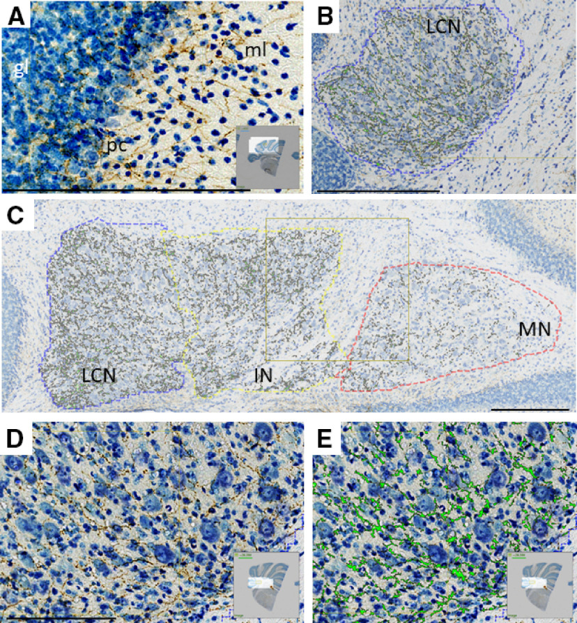
Figure 1.
Immunohistochemistry reveals staining for TH-positive fibers (brown represents DAB, anti-TH antibody; blue represents thionine/Nissl stain), which innervate multiple regions of cerebellum in coronal sections. Staining was positive in each of 20 animals. A, Microscope image of CCtx with staining for TH. ml, Molecular layer; pc, PC layer; gl, granule cell layer. B, Representative image of TH+ staining in LCN, with green overlay of pixels determined to have TH positivity by analysis software. C, Representative image of TH+ staining in CN (LCN, IN, and MN) analyzed in this study with green overlay of pixels determined to have TH positivity by analysis software. D, Higher magnification of image in B, with TH+ staining (brown) in the LCN. E, Higher magnification of image in F, with TH+ staining overlain with green pixels determined to have TH positivity by analysis software in the LCN. Scale bars: A-E, 250 μm. A, D, E, Insets, Origin of images from complete coronal section.

