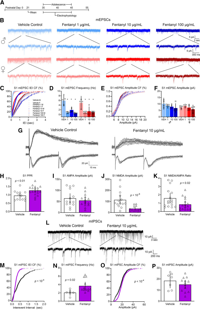
Figure 3.
Perinatal fentanyl exposure impairs synaptic transmission in somatosensory cortical neurons. A, Timeline depicting slice electrophysiology recordings in S1 layer 5 neurons of adolescent mice. B, Example traces of mEPSCs. C, Cumulative frequency plot of the interevent intervals of mEPSCs are shifted to the right in fentanyl-exposed mice compared with controls. D, Grouped data reflect decreased mEPSC frequency in fentanyl-exposed mice. There were no differences in the cumulative frequency of the mEPSC amplitude (E) nor in the grouped data for mEPSC amplitude (F). G, Example traces of evoked paired pulse and NMDAR-mediated response. Perinatal fentanyl exposure results in increased paired pulse ratio in fentanyl-exposed mice (H). There were no differences in AMPAR-mediated response amplitude (I). J, There was decreased NMDAR-mediated response amplitude, which is also reflected in the NMDA/AMPA ratio (K). L, Example traces of mIPSCs. M, Cumulative frequency plot of the interevent intervals of mIPSCs are shifted to the left in fentanyl-exposed mice compared with controls. N, Grouped data reflect increased mIPSC frequency in fentanyl-exposed mice. O, Cumulative frequency plot of the mIPSC amplitude was shifted to the right. P, There were no differences in grouped data of mIPSC amplitude. Data depict means for parametric or medians for non-parametric comparisons with 95% confidence intervals.

