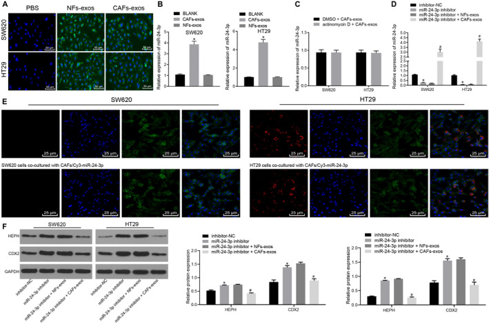
Figure 6.

miR‐24‐3p can be transferred directly from CAFs to CC cells. A, The internalization of CAFs‐exo or NFs‐exo in HT29 and SW620 cells observed under a LSCM (×200). B, Expression of miR‐24‐3p in SW620 and HT29 cells co‐cultured with CAFs‐exo or NFs‐exo measured by RT‐qPCR normalized to U6. C, The expression of miR‐24‐3p in CC cells treated with CAFs‐exo after treatment of actinomycin D determined by RT‐qPCR normalized to U6. D, The expression of miR‐24‐3p in CC cells after treatment of miR‐24‐3p inhibitor and exosomes examined by RT‐qPCR normalized to U6. E, Images of HT29 cells or SW620 cells co‐cultured with CAFs transfected with Cy3‐labelled miR‐24‐3p observed under LSCM (×400). F, The protein expression of CDX2 and HEPH in CC cells after treatment of miR‐24‐3p inhibitor and exosomes analysed by Western blot analysis normalized to GAPDH. * P < .05 vs untreated HT29 cells or SW620 cells or HT29 or SW620 cells transfected with inhibitor‐NC. # P < .05 vs HT29 or SW620 cells transfected with miR‐24‐3p inhibitor + NFs‐exo. Measurement data were expressed as mean ± standard deviation. Data between two groups were compared using unpaired t test. Comparisons among multiple groups were conducted by one‐way analysis of variance (ANOVA), followed by Bonferroni's post hoc test. The cell experiment was repeated three times
