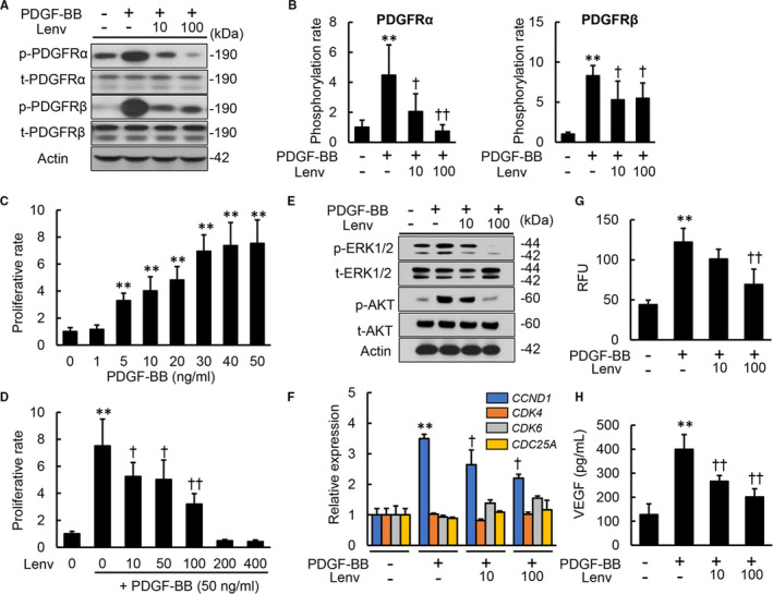
FIGURE 2.

Effects of lenvatinib on in vitro PDGF signalling pathway in LX‐2 cells. A, Western blots (WBs) of whole cell lysates for the phosphorylation of PDGFRα and PDGFRβ on LX‐2 cultured with Lenv (0‐100 nmol/L) for 2 h. B, Semi‐quantification of phosphorylation rate in PDGFRα and PDGFRβ related to Figure 2A. C, Cell proliferation of LX‐2 cells stimulated by PDGF‐BB (0‐50 ng/mL) for 24 h. D, Cell proliferation of LX‐2 cells incubated with Lenv (0‐400 nmol/L) for 24 h. E, WBs of whole cell lysates for total‐ and phospho‐ERK1/2 and AKT on LX‐2 cultured with Lenv (0‐100 nmol/L) for 6 h. F, Relative mRNA expression levels of cell cycle‐related markers in LX‐2 incubated with Lenv (0‐100 nmol/L) for 12 h. The mRNA expression levels were measured by qRT‐PCR, and GAPDH was used as internal control. G, Cell migration of LX‐2 incubated with Lenv (0‐100 nmol/L) for 6 h. Relative fluorescence units (RFU) was determined as cell migration. H, VEGFA levels in LX‐2‐cultured media. Actin was used as the loading control for WBs (A and E). Cells were pre‐treated with PDGF‐BB (50 ng/mL) 2 h before Lenv treatment (A,B,D‐H). Data are mean ± SD (n = 3 independent experiments with n = 3 (B), n = 12 (C,D,F) or n = 6 (G,H) samples per condition). Quantitative values are relatively indicated as fold changes to the values of non‐treatment groups (B‐D,F). *P < 0.05; **P < 0.01, indicating a significant difference compared with non‐treatment groups (B‐D, F‐H). † P < 0.05, †† P < 0.01, indicating a significant difference compared with PDGF‐BB(+)/Lenv(−) groups (B,D,F‐H)
