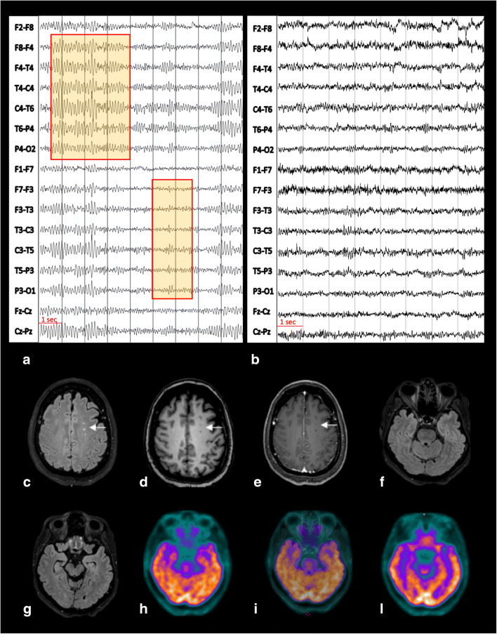
Fig. 1.
The first EEG, recorded before therapy initiation, showed a pattern of focal slow activity and spikes in the fronto-temporal area bilaterally (abnormalities magnified in red squares) (a). The second EEG, performed 4 weeks after initiation of antiseizure medication, did no longer reveal any pathological alteration (b). EEG recordings were performed with scalp electrodes placed according to the international 10–20 system with bipolar montage; brain MRI at the level of the centrum semiovale demonstrated only few hyperintense foci on FLAIR (c), mildly hypointense on T1-weighted images (d) with no contrast enhancement (CE) on corresponding CE T1-weighted images (e). These abnormalities keep with minimal non-specific changes (arrow, c–e). Co-registered FLAIR (f, g), 18F-FDG PET (h, l), and fused PET/MRI FLAIR images (i) did not show any temporal lobe abnormalities, with physiological tracer uptake according to patient’s age

