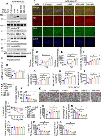
Fig. 6. Fragments of UNC5C cleaved by δ-secretase induce neuronal cell death in the hippocampus of APP/PS1 mice.

(A) WB showed the expression of UNC5C full-length, UNC5C 1–467, 1–547, 468–931, and 548–931 fragments in the hippocampus. The hippocampal lysates were detected with various indicated antibodies. Actin, loading control. Control: LV-GFP; full length: LV–GFP-UNC5C full length; 1–467: LV–GFP-UNC5C 1–467; 1–547: LV–GFP-UNC5C 1–547; 468–931: LV–GFP-UNC5C 468–931; 468–931: LV–GFP-UNC5C 468–931. (B) δ-Secretase activity in the hippocampus of above mice. Data were means ± SEM; n = 3 mice per group. (C) IF signals of ThS (green) and anti-Aβ (red) were detected in the hippocampus injected with various viruses. The nuclei were stained with DAPI. Scale bar, 30 μm. (D and E) Quantification of relative immunoreactivity was shown. (F to I) ELISA quantification of Aβ in the brain lysates of above mice. Data were means ± SEM; n = 3. (J and K) The synaptic density in the hippocampus of above mice determined by electron microscopy (means ± SEM; n = 6). Scale bar, 1 μm. (L and M) MWM analysis as time to platform (latency, s) and integrated latency (AUC) for above mice (means ± SEM; n = 7). (N and O) Probe trail and swim speed of MWM test (means ± SEM; n = 7). (P and Q) Fear conditioning test including cued fear conditioning test and contextual fear conditioning test (means ± SEM; n = 7). *P < 0.05 by one-way ANOVA followed by Tukey’s multiple-comparison test.
