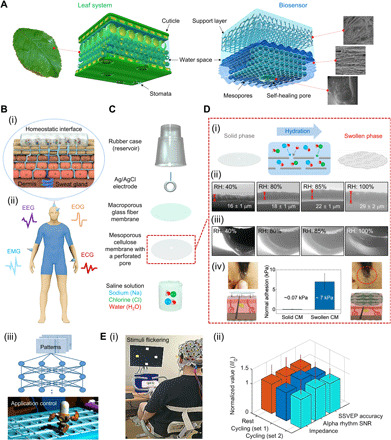
Fig. 1. Design, mechanism, and applications of cellulose-based biosensor.

(A) Structure of the proposed biosensor surface based on two types of porous membranes that mimic the leaf architecture. (B) (i) Illustration of a homeostatic interface between skin and sensor surface. (ii) Schematic illustration of measurement locations on a human model for multiple acquisition of EP signals. (iii) The flow of application control through artificial intelligence learning. (C) Exploded-view schematic illustration of a cellulose biosensor (CS). (D) (i) Schematic illustration showing the direction of ion/water permeation along the cellulose porous layers (center). (ii and iii) ESEM images of the cross section of CMs under different conditions of RH. In (iii), the perforated pore was closed by the self-healing effect of CM. (iv) Comparison of the adhesion strengths of solid and swollen CM. (E) (i) Photograph of an individual performing the steady-state visual evoked potentials paradigm while pedaling an exercise bike. (ii) Normalized comparison of the performances of CSs in resting, cycling set 1, and cycling set 2, respectively. Photo credit: Ji-Yong Kim, Korea University.
