
Fig. 3. Comparison of the number of PICs in the HEK cell membrane proteome using different digestion protocols.
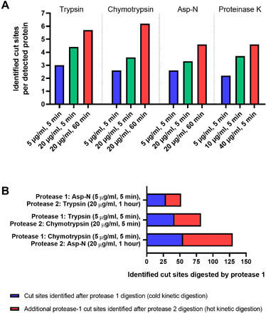
(A) Identified cut sites per detected protein with varying protease exposure. Membrane-derived vesicles from HEK cells were subjected to varying protease exposure (concentration and time) using either trypsin, chymotrypsin, Asp-N, and proteinase K. The number of identified extracellular cut sites per detected membrane protein is shown. The number of identified cut sites increases with increasing protease exposure. (B) Comparison of the number of identified cut sites after using a cold kinetic digestion (protease 1) alone or in combination with a sequential hot kinetic digestion (protease 2).