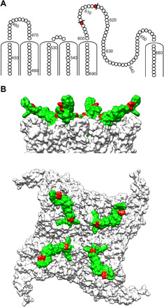
Fig. 4. Location of PICs on TRPV1 with corresponding epitope region used for development of the modality-selective anti-TRPV1 antibody OB1.

(A) Snake plot of hTRPV1 showing PICs (red) in the extracellular region, identified after a cold kinetic digestion using proteinase K. (B) Top and side view of a homology model of hTRPV1, showing the identified PICs marked in red and the epitope region translated into a synthetic antigen for development of OB1 marked in green.
