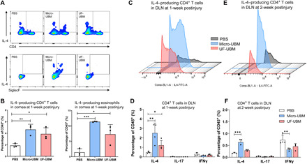
Fig. 3. Immune response in wounded corneas and draining lymph nodes (DLN).

(A) Multiparametric flow cytometry analysis of IL4+ CD4+ T cells (CD45+CD3+) and IL-4+ SiglecF+ myeloid cells (CD45+CD11b+) isolated from the wounded cornea 1 week after surgery. (B) IL-4 expression in CD4+ T cells and SiglecF+ eosinophils at 1 week after surgery (n = 3, each data point represents cells collected from five corneas). *P < 0.05, **P < 0.01, and ***P < 0.001. (C) IL-4 expression in CD4+ T cells isolated from draining lymph nodes at 1 week after surgery. (D) IL-4, IL-17, and IFNγ expression in CD4+ T cells at 1 week after surgery. (E) IL-4 expression in CD4+ T cells isolated from draining lymph nodes at 2 weeks after surgery. (F) IL-4, IL-17, and IFNγ expression in CD4+ T cells at 2 weeks after surgery. n = 4, *P < 0.05, **P < 0.01, and ***P < 0.001.
