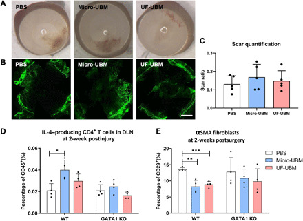
Fig. 4. Corneal scarring in Gata1−/− mice.

(A) Gross photos of wounded corneas of Gata1−/− mice treated with PBS, micro-UBM, and UF-UBM. (B) Immunostaining of α-SMA in PBS-, micro-UBM–, and UF-UBM–treated Gata1−/− corneas. Scale bar, 1 mm. (C) Scar quantification of Gata1−/− mice at 2 weeks after surgery. n = 4. (D) IL-4 expression in CD4+ T cells isolated from draining lymph nodes of wild-type (WT) and Gata1−/− mice at 2 weeks after surgery. n = 4, *P < 0.05. (E) α-SMA+ corneal fibroblast population isolated from wounded corneas of WT and Gata1−/− mice at 2 weeks after surgery. n = 4, **P < 0.01 and ***P < 0.001.
