
Figure 7.
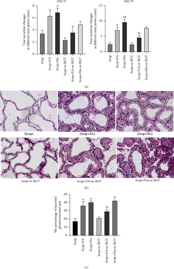
α7nAChR is involved in the protective effect of EA on the LG: (a) tear fluid flow (n = 6); (b) histopathological images of the cornea (hematoxylin-eosin staining, ×20) on day 35 (n = 3); (c) the percentage of lacrimal gland area/total area (%). Quantitative data are expressed as mean ± SEM. #P < 0.05 and ##P < 0.01 vs. the Scop group; %P < 0.05 vs. the Scop+EA group.