Figure 3.

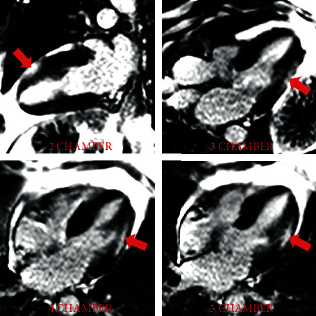
Late gadolinium enhancement (red arrows) in a midwall and epicardial distribution involving the inferolateral, anterolateral, and anterior walls consistent with myocarditis (long axes).

Late gadolinium enhancement (red arrows) in a midwall and epicardial distribution involving the inferolateral, anterolateral, and anterior walls consistent with myocarditis (long axes).