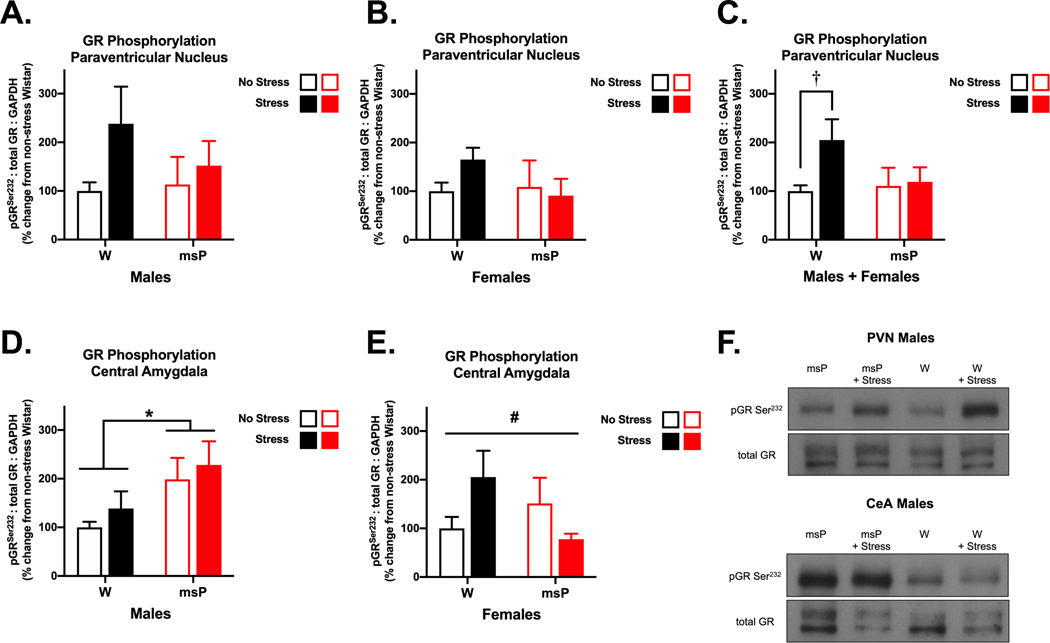
Fig. 3. Glucocorticoid receptor signaling is dysregulated in msP rats.
Male and female Wistar (W) and Marchigian Sardinian alcohol-preferring (msP) rats were examined for phosphorylation of the serine 232 site on glucocorticoid receptors (GR) in the paraventricular nucleus of the hypothalamus (PVN) (A-C) and the central nucleus of the amygdala (CeA) (D, E) following a restraint procedure or under no stress conditions (n= 6–13 per group). A combined analysis of the PVN allowed us to better discern genotypic differences across sex. Here, restraint stress induced an increase in phosphorylated GR levels in Wistar, but not msP rats. In CeA tissue, male msPs displayed an innate increase in phosphorylated GR levels that remained persistently elevated across stress conditions. Comparatively, in females, we found a significant interaction of stress and genotype, although no specific group differences emerged. Data reflect mean changes in phosphorylation levels expressed as a percentage (± SEM) of non-stressed Wistar controls. The western blot images (F) depict representative data for significant differences in phosphorylation normalized for the expression of total GR. Asterisks (*) indicate significant differences in genotype, daggers (†) indicate significant differences in stress conditions and pound signs (#) indicate a significant interaction of genotype and stress (p<0.05).

