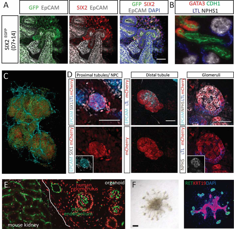
Figure 3. Cellular complexity, lineage relationships and maturation of kidney organoids.
A. The SIX2-expressing mesenchymal progenitor population within a kidney organoid generated from a SIX2-GFP fluorescent reporter iPSC line [132]. B. Nephrons within kidney organoids show patterning and segmentation into glomerulus (NPHS1, white), proximal (LTL, blue), distal (CHD1, green) and connecting (GATA3 (red) and CDH1 (green)) segments. C. Confocal imaging stack within a kidney organoid showing the presence of glomeruli comprised of podocytes (NPHS1, red) within a Bowman’s capsule of parietal epithelial cells (CLDN1, green) surrounded by an endothelial plexus (CD31; aqua). Image courtesy of Aude Dorison. D. SIX2 lineage tracing using an engineered induced pluripotent stem cell line reveals that the nephrons arise from a SIX2-expressing nephron progenitor. Initiation of SIX2 expression results in Cre-mediated excision of a GFP cassette, enabling the expression of mCherry fluorescent protein from the same locus. Lineage tracing showed that SIX2-expressing progenitors gave rise of proximal tubule, distal tubule and glomeruli within human kidney organoids [16]. E. Kidney organoid transplanted under the renal capsule of an immunocompromised mouse. After 14 days, there is clear evidence of angiogenic ingrowth of murine blood vessels (MECA32, green) forming glomerular capillaries within human organoid glomeruli. Cells of the organoid can be identified as human via staining with the human nuclear antibody (red). Image courtesy of Michelle Scurr. F. An example of a ureteric organoid generated via stepwise patterning to anterior intermediate mesoderm, subsequent isolation of nephric duct progenitors based upon surface expression of CXCR4/KIT and subsequent culture in Matrigel [76]. Ureteric organoids are shown here brightfield (left) and with immunofluorescence staining for RET and KRT19. Adapted from [76].

