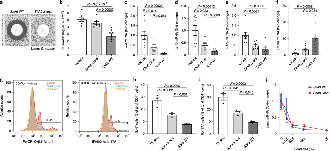
Fig. 2 |. Anti-inflammatory action of ShA9 is mediated by mechanisms independent of antimicrobial activity against S. aureus.
a, Radial diffusion assay of S. aureus growth with WT ShA9 and lantibiotic knockout mutant of ShA9 (ShA9-Δlanti). b–f, Live S. aureus recovered from lesional back skin by swab (b) and relative abundance of mRNA of Il-4 (c), Il-13 (d), Il-17a (e) and cathelicidin antimicrobial peptide in skin (f) of Flgft/ft Balb/c mice treated as in Fig. 1a–c with ShA9 WT, ShA9-Δlanti or vehicle. Data represent the mean ± s.e.m. of biological replicates from independent mice (vehicle: n = 8; ShA9-Δlanti: n = 8; ShA9 WT: n = 9). Expression of each gene was normalized to that of Gapdh (c–f). g, FACS analysis of IL-4+ CD4+ and IL-17A+ CD4+ cells isolated from the skin of mice treated as in Fig. 1a–f after twice-daily topical application of ShA9 WT, ShA9-Δlanti or vehicle for 3 d. Gating strategy for this analysis is shown in Extended Data Fig. 2. h,i, Quantification of numbers of CD4+IL-4+ (h) and CD4+IL-17A+ (i) cells shown in g. Data represent mean ± s.e.m. of biological replicates from independent mice (n = 3). j, Inhibition of psmα mRNA expression by S. aureus (USA300Lac) in culture after exposure to indicated dilutions of conditioned medium (CM) from ShA9 WT or ShA9-Δlanti for 18 h. USA300Lac was resistant to antimicrobial activity from ShA9. Data were normalized against the gyrB gene. Data represent mean ± s.e.m. of technical replicates from individual bacterial cultures (n = 3). The P value was calculated using one-way ANOVA (b–f, h and i). No significant difference was found using the two-tailed Mann–Whitney U-test (P > 0.05) (j).

