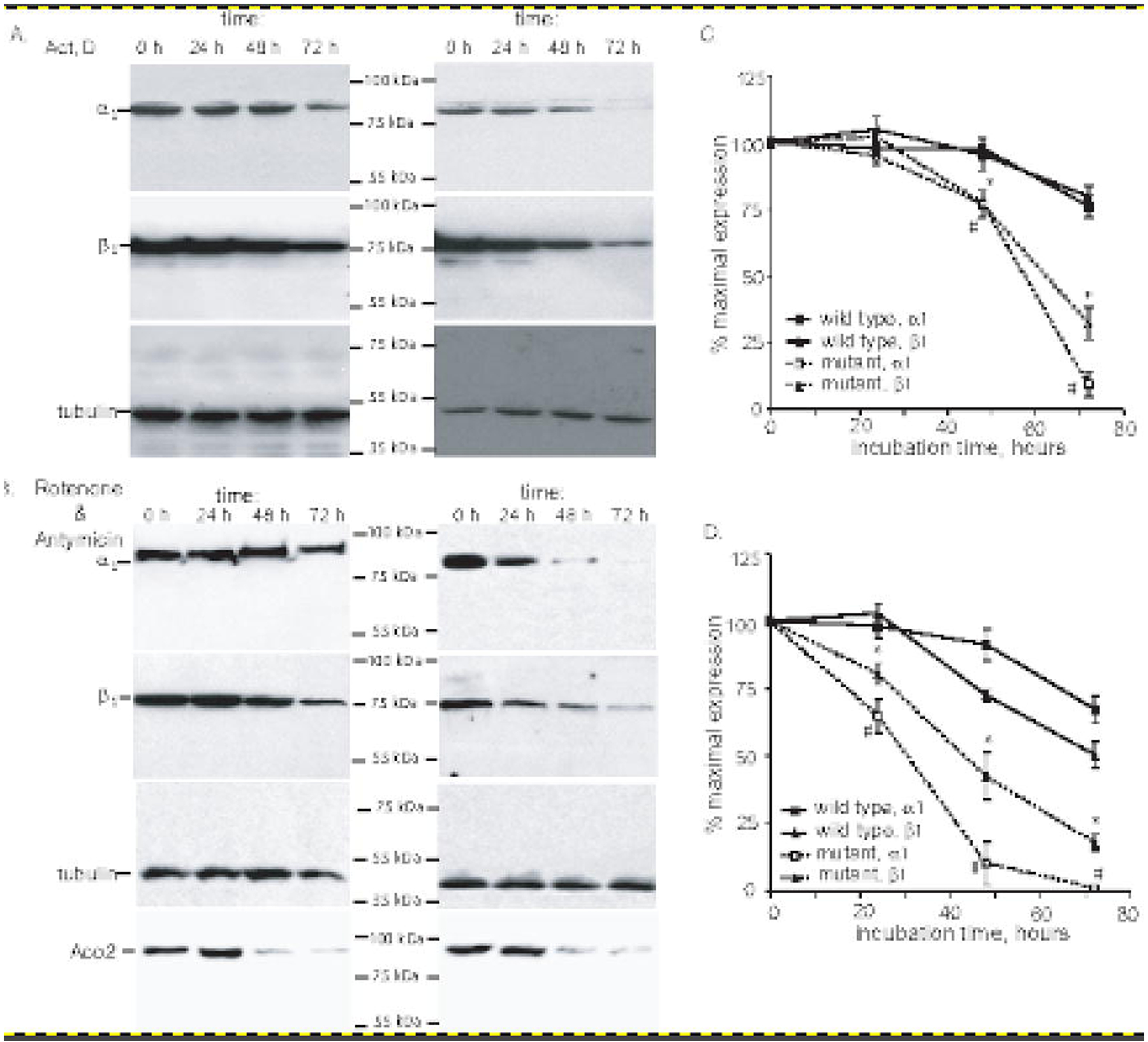
Figure 5. Diminished stability of α1Cys517Tyrβ1 sGC in COS7 cells.

(A-B): COS-7 cells were transfected with plasmids expressing wild type or α1Cys517Tyrβ1 sGC. 48 hours post-transfection, the cells were treated with 1 μM actinomicyn D (A) or 1 μM rotenone and 1 μM antimycin (B). At indicated time points, the cells were lysed and 100,000xg supernatant were used for the detection of α1, β1 and β-tubulin by western blotting. Changes in the level of ROS sensitive mitochondrial aconitase (ACO2) was evaluated in 20,000xg pellets. Shown are representative blots of three independent transfections with similar results. (C-D): Densitometry results of western blots from (A-B) showing changes in the amount of α1 and β1 subunits in COS-7 cells expressing wild type or α1Cys517Tyrβ1 after actinomycin D (A) or rotenone+antimycin (B) treatments. Signals for α1 and β1 subunits are normalized to β-tubulin signal. Data are presented as % of signal obtained in control samples, before the exposure to actinomycin D (A) or rotenone+antimycin (B). Data are mean ± SD from three independent transfections.
