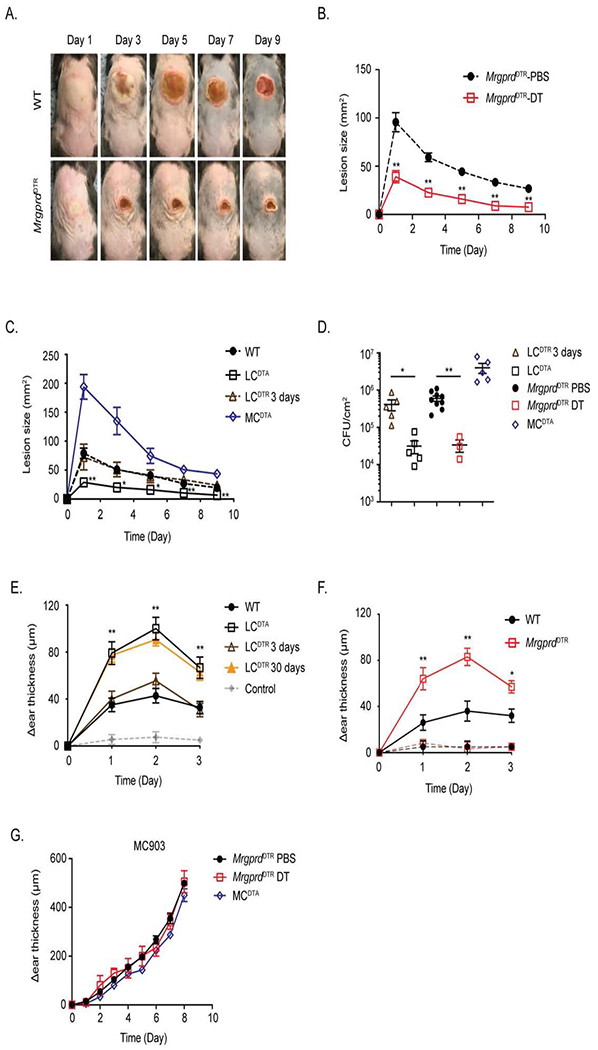
Figure 3. Ablation of MrgprD-expressing neurons or LC augments S. aureus host defense and CHS.

(A) Representative images of skin lesions from PBS (WT) or DT treated MrgprdDTR mice on indicated day after i.d. inoculation with 107 S. aureus. (B) Skin lesion area calculated using ImageJ from PBS or DT-treated MrgprdDTR mice at the indicated time is shown. (C) As in B, skin lesion area is shown at the indicated time for WT, LCDTA, 3-day DT-treated LCDTR and MCDTA mice. (D) Quantification of CFU isolated from infected skin tissue at Day 10 after infection from mice in B and C. (E) WT, LCDTA, 3-day or 30-day DT-treated LCDTR and MCDTA mice were sensitized with 0.25% DNFB on shaved abdomens on day 0, followed 5 days later with a 0.2% DNFB challenge on the ear. The data shown represent the change in ear thickness at the indicated time with the thickness prior to challenge. The change in ear thickness of all mice sensitized with vehicle was merged into grey line. (F) As in E, comparing DNFB CHS responses sensitized with 0.25% DNFB (thick lines) or vehicle alone (thin lines) in PBS or DT-treated MrgprdDTR mice. (G) The ears of PBS or DT treated MrgprdDTR and MCDTA mice were daily challenged with MC903 and monitored for ear swelling. Results are represented as mean ± SEM from two independent experiments using cohorts of 4-7 mice each for B, C, E, F and G. Each symbol in D represents data from an individual animal. Significance was calculated using unpaired Student’s t test to compare with WT group. *p < 0.05, **p < 0.01.
