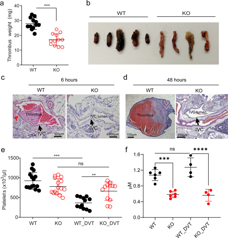
Fig. 7. Mfsd2b knockout mice show reduced deep vein thrombosis.
a, b Global Mfsd2b knockout mice had reduced venous thrombosis in IVC stenosis model. Weights (a) and representative images (b) of thrombi collected 48 h post stenosis of IVC from WT and KO, respectively. Data are mean and SD. Each dot represents one animal (n = 12). ***P < 0.001. Two-tailed unpaired t-test was used. WT wild-type, KO knockout. c, d Representative histology of inferior vena cava (IVC) after 6 and 48 h of stenosis in WT and Mfsd2b KO mice. Experiments were performed at least twice (n = 2–3). Arrows indicate IVC. e Thrombosis resulted in reduced platelets count in blood of WT mice. Data are mean and SD. Each dot represents one animal (n = 15 for WT and KO, n = 12 for WT-DVT and KO-DVT). **P < 0.01, ***P < 0.001; ns not significant. One-way ANOVA was used. f Venous thrombosis did not induce the loss of total plasma S1P. Plasma S1P from indicated mice was collected before and 48 h after DVT. Note that the analysis of S1P for plasma collected from DVT experiments was acquired at the same time with the data shown in Fig. 4e. Thus, data for S1P levels from WT and global Mfsd2b KO were used for comparison. Data are mean and SD. Each dot represents one animal (n = 4–6). ***P < 0.001; ****P < 0.0001; ns not significant. One-way ANOVA was used. DVT deep vein thrombosis.

