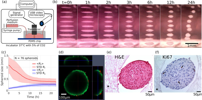
Figure 1.
Self-organization of MSCs cells sheets into spheroid shapes and biological viability. (a) Presentation of the experimental setup. A computer drives the wave generation and the acquisition of images. The transducer converts the electrical energy into acoustic energy inside the chip cavity and produced the acoustic levitation of cells over multiple acoustic pressure nodes. A USB digital microscope took side-view pictures and time-lapses of the cell sheets in levitation and self-organization into spheroids. A syringe pump allows a continuous perfusion of the cell medium during the 24 h acoustic manipulation. The whole setup, apart from the computer and the generator, was put in an incubator for a well-controlled cell culture environment. (b) Time lapse snapshots of the cell self-organization. The whole time-lapse is available on the Supplementary Video 2. At the beginning, the cells were trapped in monolayers. Then, the cells sheets contracted and the cells self-organized to reach a stable spheroid shape. (c) Time-evolution of the axial and the radial dimensions study of the cells aggregates, averaged over 76 individuals, computed from the time-lapse observations. At , the widths ranged from 0.75 to 1.5 mm depending on the aggregate location. However, all the heights were similar because of the monolayer shape. After 15 h of levitation, every MSC aggregates have reached a stable spheroid shape (). (d) Confocal and fluorescence imagery of a spheroid just after the acoustic levitation. A live/dead kit was used to label the cells. Because of the limited optical penetration or the substrate diffusion, only the cells at the surface were visible and a majority of them showed a living signal. (e) Histology of MSC spheroids. After a 24 h levitation, spheroids were collected from the chip and were immediately fixed in parafolmaldehyde 4% overnight. The spheroids were then embedded in fibrin network (described by the symbol * on the images) for an easy handling of paraffin inclusion and histological preparation. Spheroids were stained with H&E for general structure evaluation and (f) Ki67 for proliferation.

