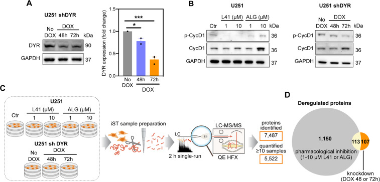
Fig. 1. Proteomic characterization of DYRK1A inhibition in U251 glioblastoma cells.
A DYRK1A (DYR) expression in U251 cells transfected with doxycycline (DOX)-inducible shRNA targeting DYRK1A (U251 shDYR). Representative images and densitometric quantification of three independent experiments are shown (mean ± SEM, unpaired t-test, *P < 0.05; ***P < 0.001). B Expression of phosphorylated (p-CycD1) and total cyclin D1 (CycD1) in U251 cells treated ± DYRK1A inhibitors L41 and ALGERNON (ALG) for 72 h, and in U251 shDYR cells treated ± DOX. Representative images of two independent experiments are shown. C Design and summary of the proteomic analysis of U251 cells treated ± L41 and ALG for 72 h, and U251 shDYR cells treated ± DOX. D Venn diagram showing overlap among proteins deregulated after DYRK1A pharmacological inhibition (1263 proteins total) and genetic knockdown (220 proteins total).

