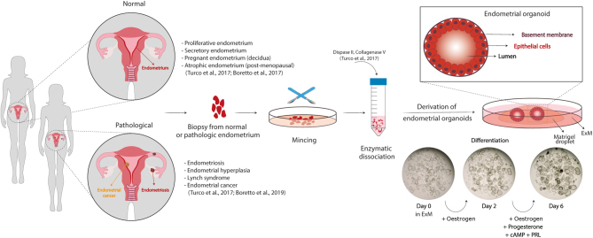
Figure 1.
Organoids of the human endometrium. Organoids can be derived from tissue biopsies from non-pregnant women in the proliferative or secretory phase of the menstrual cycle or from pregnant and postmenopausal women. The sample is minced mechanically and enzymatically to obtain epithelial isolates which are resuspended in an extracellular matrix (Matrigel) to allow self-organization of the organoids into a cystic structure formed by columnar epithelial cells lining a central lumen. They can be propagated long-term in a chemically defined medium (Expansion medium, ExM) containing nicotinamide, supplements (N2, B27), activators of WNT signalling (RSPO1 and CHIR99021), growth factors (EGF, HGF, and FGF10) and inhibitors of TGFB and BMP signalling pathways (A83-01 and Noggin, respectively). They recapitulate the morphological, molecular and functional features of endometrial glands in vivo and respond to steroid hormones, oestrogen and progesterone. To induce differentiation, they are primed with oestrogen for 2 days and then treated with a combination of oestrogen, progesterone, prolactin and cAMP for another 4 days. Brightfield images depict morphological changes in an organoid culture before (day 0), during (day 2) and after (day 6) their differentiation with hormones. Organoids with thicker epithelia and secretions in the lumen are visible after full hormonal stimulation. Organoids can also be derived from pathological endometrium including endometriosis, endometrial hyperplasia and cancer.

 This work is licensed under a