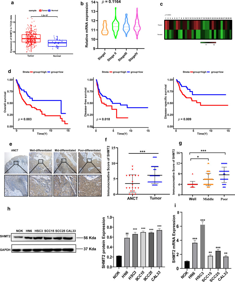
Fig. 1.
SHMT2 was overexpressed in OSCC tissues and predicted poor prognosis. a Relative analysis of SHMT2 mRNA in OSCC (n = 313) and adjacent normal tissues (n = 30) from the TCGA database. b Analysis of SHMT2 mRNA level in different pathologic stages of OSCC samples in the TCAG database (stage I, n = 19; stage II, n = 54; stage III, n = 56; stage IV, n = 162). c SHMT2 expression level in 30 paired oral tumor tissues and adjacent normal tissues in the TCGA database. d Kaplan–Meier survival curves for overall survival (n = 312), disease free survival (n = 312) and disease specific survival (n = 295) in OSCC patients with high and low expression of SHMT2. e Immunohistochemistry staining of SHMT2 in ANCT and tumors with different degrees of differentiation. Magnification of 100 × (up) and 400 × (down). f Immunohistochemical staining score of SHMT2 in ANCT (n = 19) and OSCC (n = 91) tissues. g Immunohistochemical staining score of SHMT2 in three different differentiation degrees of OSCC (Well: n = 17, middle: n = 41, poor: n = 33). h Western blot pictures and quantitative analysis of SHMT2 in NOK and TSCC cell lines. i Real time PCR analysis of SHMT2 in NOK and TSCC cell lines. P-values were obtained using Student's t-tests, one-way ANOVA tests, and log-rank tests. All data are shown as mean ± SD. * p < 0.05, ** p < 0.01, *** p < 0.001. OSCC, oral squamous cell carcinoma; TSCC, tongue squamous cell carcinoma ANCT, adjacent noncancerous tissue samples; NOK, normal oral keratinocyte

