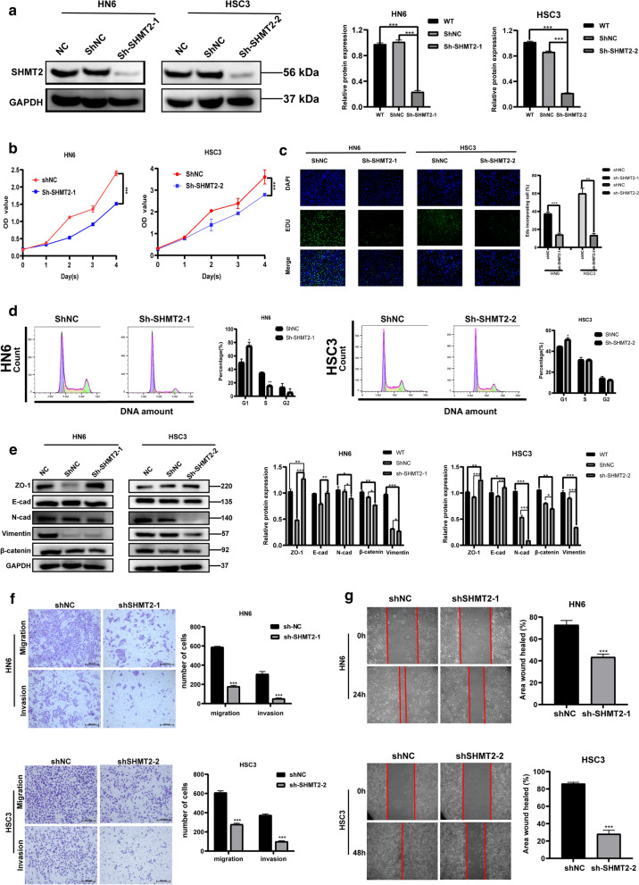
Fig. 6.
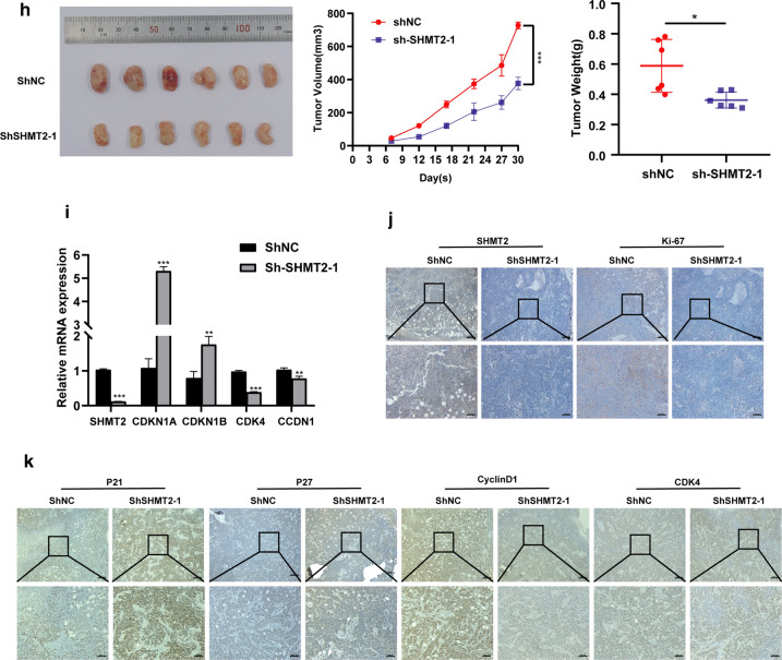
Silencing SHMT2 inhibited the invasive, migrate ability and EMT, and suppressed TSCC cell growth in vitro and in vivo. a Western blot analysis and quantitative calculation for SHMT2 level in HN6 and HSC3 cells after lentiviral transfection. b Cell viability for HN6 and HSC3 were detected by CCK8 assay. c Images of EdU staining in sh-SHMT2 and sh-NC group (left), and quantitative analysis of EdU stained cells in HN6 and HSC3 (right). d Flow cytometric analysis of sh-RNA treated cells. e Western blot analysis confirmed that silenced SHMT2 expression inhibited EMT of TSCC cells. f Representative photos of trans-well assay in HN6 and HSC3 cells after treated with shRNA (left), and quantification of cell numbers in HN6 and HSC3 (right). g Representative photos of wound healing assay in HN6 and HSC3 cells after treated with shRNA (left), and quantification of cell numbers in HN6 and HSC3 (right). h Image for tumors
taken from mice injected with sh-NC and sh-SHMT2 HN6 cells; Tumor volume growth curve of mice; Tumor weight was measured after mice were sacrificed. i Real time PCR was used to evaluate expression of SHMT2, CDKN1A, CDKN1B, CCDN1 and CDK4 in mouse tumors. j, k Immunohistochemistry staining was performed to analyze protein expression of SHMT2, Ki-67, P21, P27, cyclinD1, and CDK4 in tumor tissue specimens from sh-SHTMT2 and control groups of mice. Magnification at 50 × (up) and 100 × (down). P-values were obtained by Student's t-tests and two-way ANOVA tests * p < 0.05, ** p < 0.01, *** p < 0.001. Sh, short hairpin RNA


