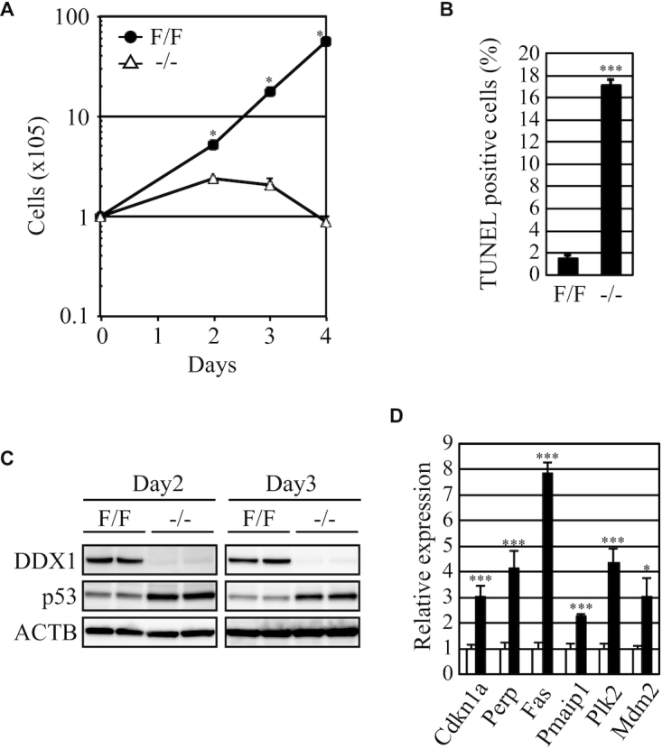
Figure 3.

Proliferation of Ddx1−/− ESCs. (A) The numbers of Ddx1F/F or Ddx1−/− cells were counted on the indicated days after sorting (n = 3; *P < 0.05). (B) Apoptosis of Ddx1F/F and Ddx1−/− cells was analyzed on day 3 (n = 3; ***P < 0.005). (C) The p53 expression levels in Ddx1F/F or Ddx1−/− cells were analyzed via western blotting. (D) The expression levels of p53-regulated genes were analyzed via qRT-PCR (n = 3; *P < 0.05; ***P < 0.005).
