Figure 3.
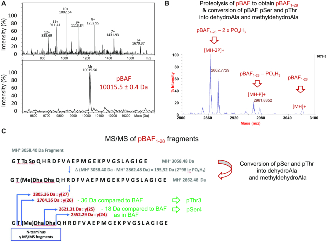
MS analyses confirm that pBAF is phosphorylated at Thr3 and Ser4. (A) Accurate mass measurement of pBAF obtained by μLC-ESI-MS. This analysis specified that the mass of pBAF is 10015.4 ± 0.6 Da. BAF did not give any multi-charged mass spectrum that could enable the measurement of its accurate molecular weight. (B, C) Identification of the phosphorylated residues by fragmentation and MS. Peptide Mass Fingerprint of pBAF digested by endo-GluC was obtained using MALDI-TOF MS in reflector positive mode. M/z peaks were then selected for MSMS sequencing by PSD-MALDI-TOFTOF. (B) Zoom view of the MSMS spectrum of the double phosphorylated BAF1–28 peptide at m/z 3058.40. Fragmentation of pBAF1–28 lead to the conversion of pSer and pThr into dehydroAla and methyldehydroAla, due to a β-elimination of the phospho-groups (56,57). (C) Further fragmentation of pBAF1–28 into y(i) peptides (containing ‘i’ amino acids) revealed that Thr3 and Ser4 are phosphorylated. The whole MS spectrum of fragmented pBAF1–28 is shown in Supplementary Figure S2B.

