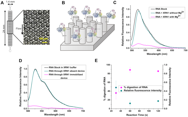
Figure 2.
Solid-phase digestion reactions of XRN1. (A) Top down view of the pillared IMER channel. (B) Schematic representation of the covalently attached enzyme on the micropillars of the device. Fluorescence emission spectra of SYTO RNASelect Green labeled monophosphorylated RNA solutions digested by XRN1 in (C) free solution and (D) Immobilized state. The reaction time was 60 s and 2.32 pmol of enzyme was used in both free solution and immobilized digestion. SYTO RNASelect Green was added after digestion and fluorescence emission spectra were taken from 495 to 700 nm with 480 nm excitation. (E) Percentage digestion and relative fluorescence intensity of digested RNA with varied reaction time and constant surface enzyme density. The XRN1 reactions were all performed at room temperature. The error bars represent standard deviations in the measurements (n ≥ 3).

