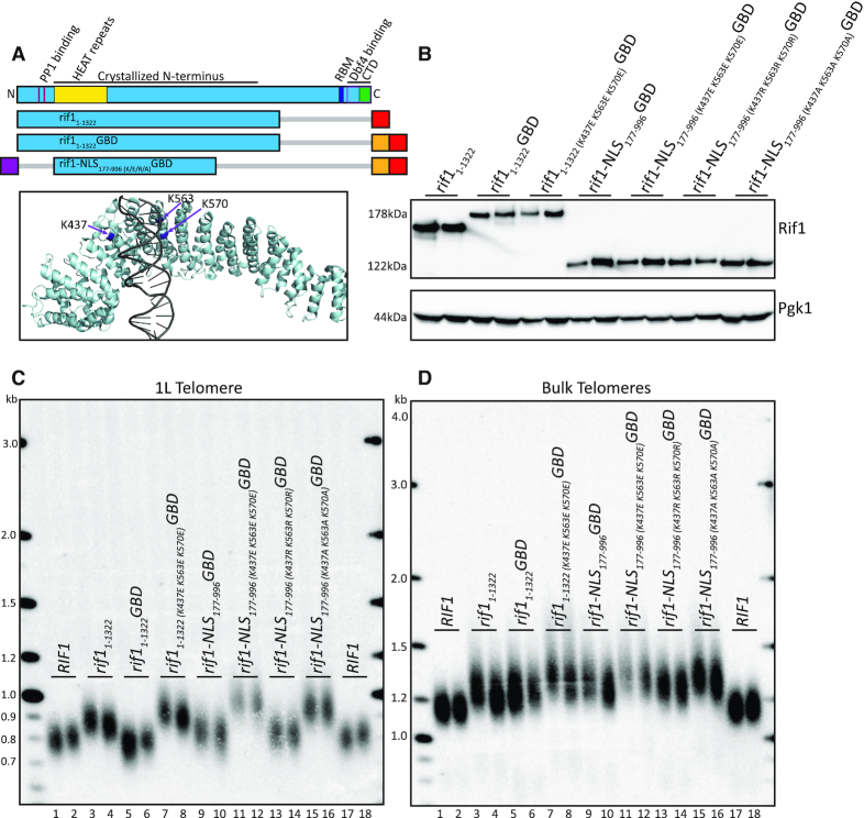
Figure 5.
Positively charged residues in HEAT repeats are required for Rif1 function when localized to telomere. (A) Rif1 domain map as in Figure 1A. Schematic of constructs tested in this figure (Blue: Rif1; Red: 6xFLAG; Orange: GBD; Purple: c-myc NLS; Gray bar: N- or C-terminal truncation of residues). PyMOL rendering of Rif1 structure with lysine residues (K437, K563, K570) depicted in Purple (PDB: 5NW5, showing one Rif1 monomer and DNA). (B) Western blot showing Rif1 (anti-FLAG antibody) and Pgk1 (anti-Pgk1 antibody, control) protein levels of indicated strains. (C) Southern blot showing 1L telomere probe for the indicated strains. (D) Southern blot from C, rehybridized with a Y’ probe to visualize ‘bulk’ XhoI restriction fragments (see ‘Materials and Methods’ section).

