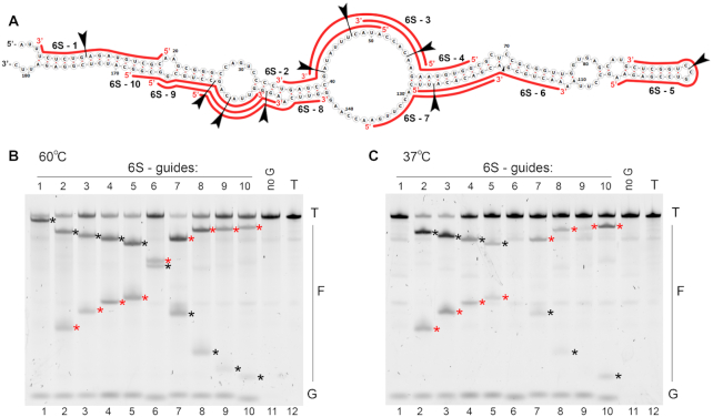
Figure 6.
RNA probing with KmAgo. (A) Secondary structure of 6S RNA used in the experiments. Positions of small guide DNA loaded into KmAgo are shown with red lines (see Supplementary Table S1 for full guide sequences). Positions of the expected cleavage sites for each guide smDNA are indicated with arrowheads. (B) Analysis of the cleavage products obtained after incubation of KmAgo with 6S RNA for 30 min at 60°C. (C) The same experiment performed at 37°C. Representative gels from two independent experiments are shown. Positions of the 5′-terminal and 3′-terminal cleavage fragments are indicated with red and black asterisks, respectively.

