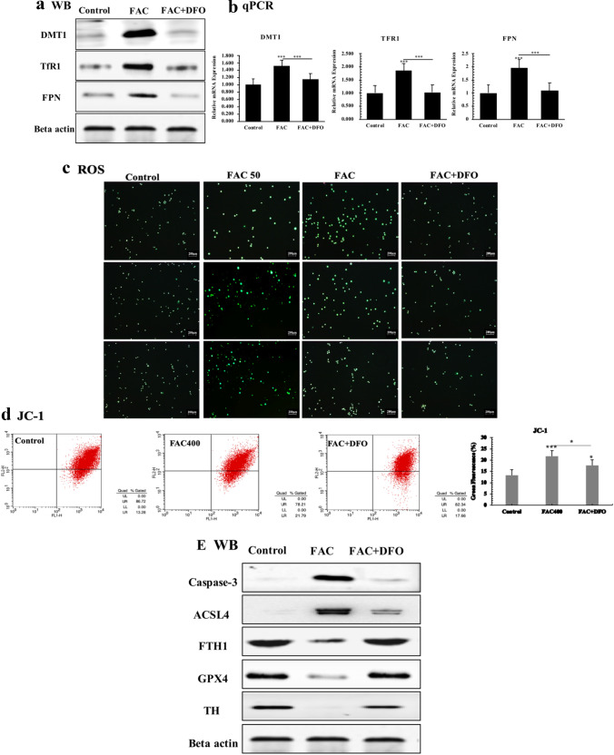
Fig. 4.
DFO rescues FAC induced iron overload. a Western blot; beta actin was used as an internal reference. b The real-time quantitative PCR analysis. DFO rescues FAC induced oxidative stress. c The fluorescence intensity of each group of cells stained with DCFH-DA was observed under a fluorescence microscope. The relative fluorescence intensity was calculated by Image J software, and the green fluorescence enhancement represented an increase in intracellular ROS. The scale was 200 μm. d Flow cytometry was used to detect the relative intensity of green fluorescence after treatment wih JC-1 of each group. DFO rescues FAC activated ferroptosis. e Western blot of ferroptosis-related proteins in PC12-NGF cells under FAC stimulation. All experiments were repeated three times and averaged. The Bonferroni method and Dunnett’s T3 method were used. The test level is α = 0.05; *P < 0.05; **P < 0.01; **P < 0.001, vs Control

