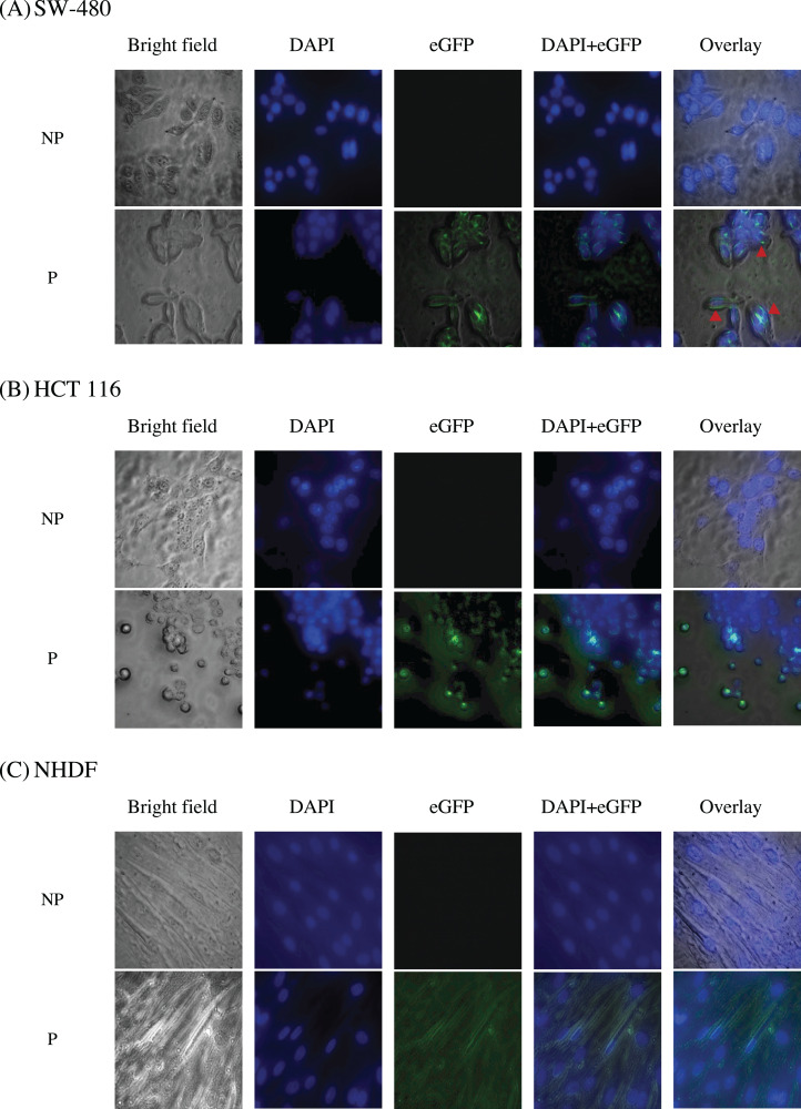
Figure 10. Immunofluorescence staining of (A) SW-480 (G12V KRAS-positive), (B) HCT 116 (G13D KRAS-positive) and (C) NHDF (wt KRAS-negative) cells.
Blue fluorescence denotes nuclei stained with DAPI, while green fluorescence represents the binding localization of scFv via eGFP. Images were obtained on an Axio Vert A1 under 630× magnification and arranged with ZEISS Zen software. Red arrows indicate scFv binding. NP, non-permeabilization; P, permeabilization.

