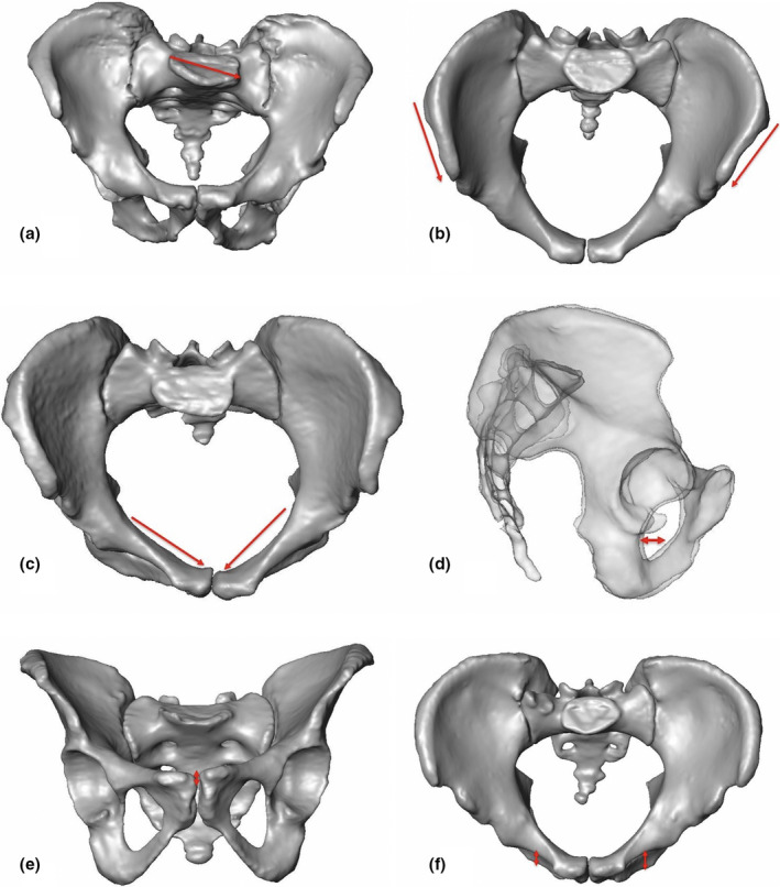
FIGURE 4.

Given 3D CT models to display the six APRs with their locations: (a) 88 years old Asian female with sacral asymmetry: 3D CT model in anteroposterior view with asymmetric oriented sacrum and lateralized and oblique sacral basis (case p457). (b) 3D CT model of a 27 years old Asian male: Inlet view demonstrates iliac crest asymmetry (asymmetric position of left versus right ASIS (case p415)). (c) 3D CT model of a 54 years old European female: Inlet view exhibits asymmetric pelvic brim (case p601). (d) Semi‐transparent lateral view of a 65 years old European female with asymmetric acetabula (case p634). (e) 3D CT model of an 86 years old European female: Outlet view with asymmetric pubic symphysis (case p526). (f) 3D CT model of a 62 years old Asian male: Inlet view with asymmetric inferior pubic ramus (case p424)
