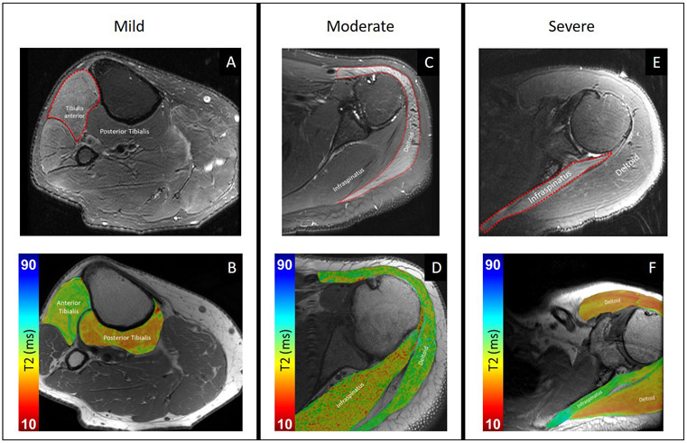
Figure 1:
Morphological, Dixon fat-suppressed T2-weighted images (top row) and T2 maps (bottom row), superimposed on proton-density weighted images, for patients with varying edema pattern severity by morphological grading. (A) 72-year-old man with recent onset right foot drop status post lumbar laminectomy two weeks prior, with mild edema pattern of the tibialis anterior muscle and normal signal intensity arising from the posterior tibialis. (B) Corresponding representative axial T2 mapping image demonstrates elevated T2 of the anterior tibialis. (C) 42-year-old man with weakness of shoulder abduction status post glenohumeral capsular repair surgery 1.5 years prior, with moderate edema pattern of all three deltoid muscle heads and normal signal intensity of the infraspinatus. (D) Corresponding representative axial T2 mapping image demonstrates elevated T2 of the deltoid. (E) 54-year-old man with spontaneous onset of severe pain followed by left shoulder abduction weakness, 8 months prior and attributed to neuralgic amyotrophy, with severe edema pattern of the infraspinatus muscle and normal signal intensity of the deltoid. (F) Corresponding representative axial T2 mapping image demonstrates elevated T2 of the infraspinatus.

