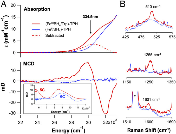
Fig. 1.
(A) Absorption (Top) and 7 T, 5 K MCD (Bottom) spectra for pterin-bound FeII-TPH (125 μM, blue) and pterin- and tryptophan-bound ternary FeII-TPH (125 μM, red). (Inset, Bottom) Near-infrared MCD spectra demonstrating the formation of a 5C site upon tryptophan binding (red) to the (FeII/BH4)-Trp active site. (B) Resonance Raman data collected at 334.5 nm (arrow in A) showing resonance enhancement of three vibrations upon tryptophan binding (0.5 mM, red) to the (FeII/BH4)-TPH active site (0.5 mM, blue).

