Fig. 2.
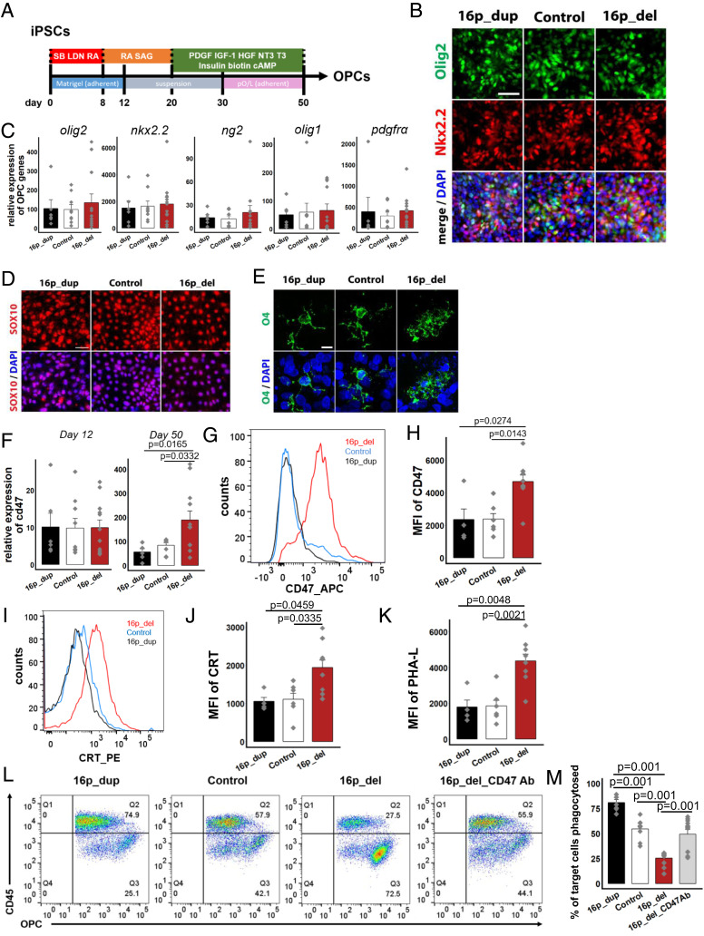
CD47 expression is increased on 16p11.2 deletion OPCs protecting them from calreticulin-mediated phagocytosis.(A) Schematic of directed differentiation of iPSCs into oligodendrocyte progenitor cells (OPCs). (B) After 12 days of differentiation, immunostaining for pre-OPC markers Olig2 and Nkx2.2. Nuclei (DAPI, blue). (C) Following 50 days of directed differentiation, quantitative RT-PCR of mRNA expression levels of genes associated with the oligodendrocyte lineage. Data represent fold change relative to undifferentiated control human iPSCs. P values not significant at 5% level. (D) Immunostaining for Sox10 after 50 days. (E) Confocal imaging showing expression of the OPC marker O4 after 50 days. (F) Quantitative RT-PCR of mRNA expression levels of cd47 after 12 days (Left) and 50 days (Right) of directed differentiation. Data represent fold change relative to undifferentiated control human iPSCs. (G) Flow cytometry histograms of the mean fluorescence intensities (MFI) of CD47 in OPCs after 50 d of directed differentiation. (H) Quantification of CD47 MFI. (I) Histograms of the MFI of cell-surface expression of CRT in OPCs. (J)Quantification of CRT MFI. (K) Quantification of PHA-L in OPCs. (L) Flow cytometry phagocytosis plots showing rates of engulfment of OPCs when cocultured with human derived macrophages (labeled with a CD45 antibody). 16p_del_CD47 Ab, OPCs differentiated from 16p_del lines pretreated with CD47 blocking antibody prior to phagocytosis assessment. (M) Percentage of phagocytosed OPCs. All data are mean ± SE. P values determined by one-way ANOVA followed by post-hoc Tukey HSD Test (C, F, H, J, K, M). n = 3 (C, F, M) and n = 2 biological replicates (H, J, K) per cell line (16p_dup, n = 2; control, n = 3; 16p_del, n = 4 cell lines). [Scale bar, 50 µm (B, D, E).]

