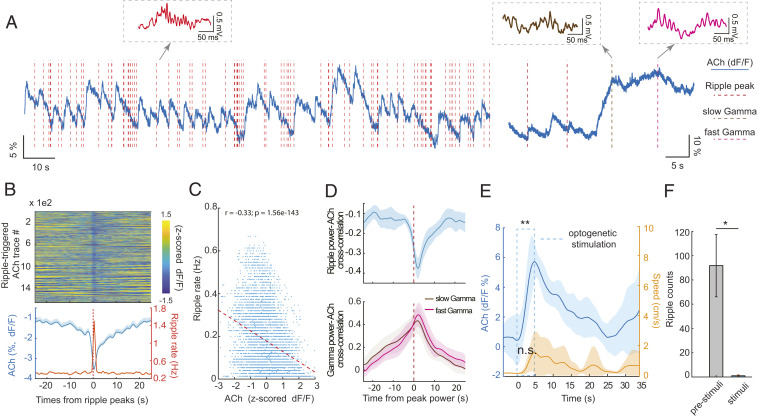
Fig. 3.
SPW-Rs occur when hippocampal cholinergic activation transiently decreases during slow wave sleep (SWS). (A) Example traces of ACh3.0 signal fluctuation (blue line) and SPW-R occurrence (red vertical dashed lines) during non-REM (Left) and non-REM–waking transient (Right). Examples of SPW-R, slow (brown) and fast gamma epochs (purple), extracted from the simultaneously recorded LFP, are shown as insets. (B, Top) Color-coded, normalized change of ACh3.0 signal surrounding SPW-Rs during non-REM sleep (n > 1,600 SPW-Rs from 4 non-REM sessions, single mouse). (B, Bottom) Mean change (± SEM) of ACh3.0 (blue) and ripple rate (orange) centered on the peak power timing of ripple events. (C) The relationship between ACh3.0 signal and ripple rate (n = 10 sessions in four mice). (D, Top) Cross-correlations between ACh3.0 signal and continuous ripple-band–filtered (140 to 250 Hz) LFP power in 0.5 s epochs (from six non-REM sleep sessions in three mice). (D, Bottom) Cross-correlation between ACh3.0 signal and continuous gamma-band–filtered (30 to 80 Hz slow gamma and 80 to 120 Hz fast gamma) LFP power (from six waking sessions in four mice). (E) Time course of optogenetically induced ACh3.0 signal during sleep. Medial septum was optogenetically stimulated by 5 s long pulses during non-REM sleep. ACh3.0 signal increased significantly during stimulation (0.48 ± 1.4% prior to stimulation; 5.95 ± 0. 83% by the end of stimulation; **P = 0.004, two-tail paired test, n = 7 trials). Movement (Right y axis) occurred on some trials, but overall, it did not change significantly during stimulation (P = 0.052, two-tail paired t test). (F) Optogenetic stimulation of cholinergic medial septal neurons suppressed SPW-R occurrence during non-REM sleep (paired t test: *P = 0.04, n = 4 mice). *P < 0.05.

