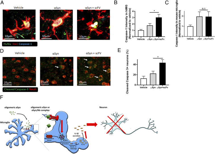
Fig. 4.
Ab–αSyn complex activates the inflammasome and induces neuronal death in hiMG-engrafted mice. (A) Representative images showing cleaved/activated caspase-1 staining in transplanted hiMG: hiMG only (Left), hiMG with αSyn (Middle), or hiMG with αSyn and Ab scFv (Right). Human nuclear antigen (HuNu; green), Iba1 (red), and activated caspase-1 (cyan). (Scale bars, 10 µm.) (B) Quantification of caspase-1 intensity in engrafted hiMG (Iba1+HuNu+ cells) (n = 6 per group). (C) Quantification of caspase-1 intensity in endogenous mouse microglia (Iba1+/HuNu− cells) (n = 6 per group). (D) Representative images of cleaved/activated caspase-3 staining in endogenous mouse neurons after transplantation of hiMG only (Left), hiMG with αSyn (Middle), or hiMG with αSyn plus scFv (Right). Cleaved caspase-3 (green, indicated by arrows) and NeuN (red). (Scale bars, 25 µm.) (E) Quantification of cleaved caspase-3 in neurons (NeuN+ cells) (n = 6 per group). (F) Schema showing the effect of αSyn on NLRP3 inflammasome activation. Oligomeric/fibrillar αSyn is released from DA neurons, and more so from A53T DA neurons. Oligomeric/fibrillar αSyn activates the NLRP3 inflammasome by providing two triggers: Stimulation of TLR2 provides the priming trigger, and induction of mitochondrial damage and/or ROS production provides the secondary trigger. This leads to the assembly of the NLRP3 inflammasome, which results in secretion of IL-1β and caspase-1, and contributes to caspase-3–associated neuronal cell death. Ab–αSyn complexes enhance inflammasome-mediated IL-1β secretion in a TLR2-dependent manner. Red arrows indicate increased response to the Ab–αSyn complexes. Graphs indicate mean ± SEM. Statistical analysis was performed using a two-way ANOVA with Bonferroni post hoc test (*P < 0.05; N.S., not significant, P > 0.9).

