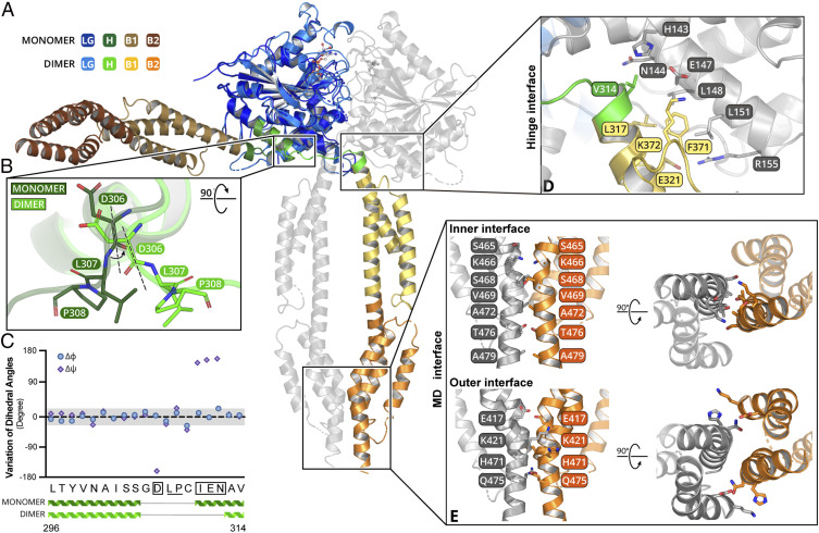
Fig. 3.
The hGBP5 MD undergoes drastic movement and forms an extended dimer interface with GTP. (A) Superimposition of nucleotide-free and nucleotide-bound structures of hGBP5 reveals a drastic rearrangement of the MD. The color scheme is shown on the left. (B) The conformational change is due to a rotation in the hinge region. D306, L307, and P308 are shown as sticks. (C) Changes of the Cα dihedral angles of residues at the hinge region are plotted. (D) The hinge interface is shown in detail. The residues involved are shown as sticks. (E) The stalk interface can be divided into an inner interface and an outer interface. The residues involved are shown as sticks.

