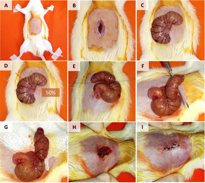
Figure 1. Cecal Ligation and Puncture Model in Rats.
A. Disinfection and shaving of the lower abdominal region. B. Longitudinal incision in the skin and muscle fascia. C. Cecum exposure. D-E. Cecal ligation (in the mid-cecum). F. Puncture of the distal cecum with a 16 G needle. G. Squeezing and distribution of feces around the cecum and peritoneal cavity. H-I. Closure of the abdominal muscle fascia and skin.

