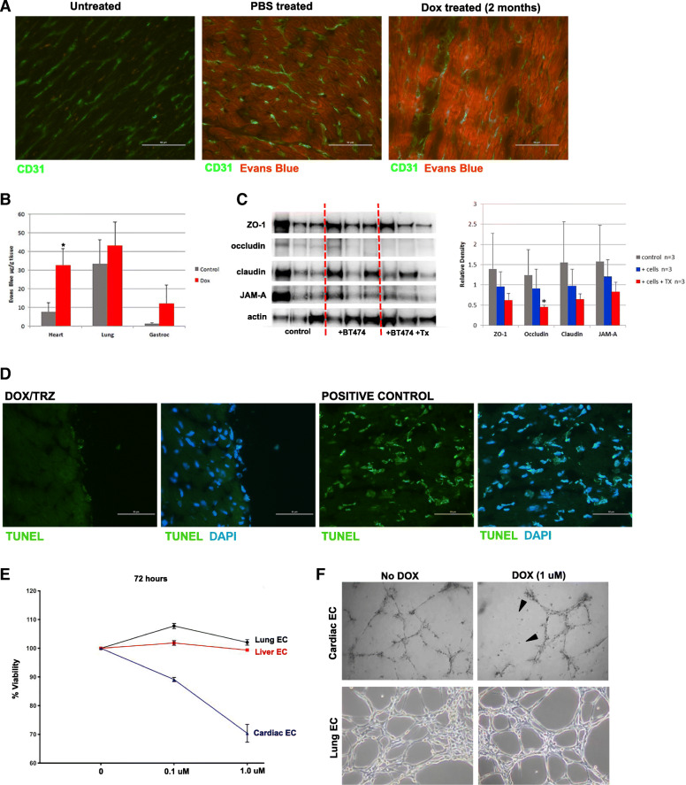
Fig. 4.
Cardiac endothelial cells (ECs) are sensitive to DOX. a Representative images of heart sections immunostained with the EC marker CD31 (green) before or after intraperitoneal injection with Evans Blue (EB) dye to assess vascular permeability with EB dye visible as red fluorescence. b miles Assay of EB extracted from labeled tissues shows 4 times more dye in hearts, but not lung or gastrocnemius muscle, from DOX-treated mice, compared to untreated. *p < 0.05. c Western blot analysis of junctional proteins in hearts from control mice (no tumor), tumor-bearing mice (+ BT474) or tumor bearing-mice treated with 5 mg/kg DOX and 4 mg/kg TRZ (+BT474 + Tx). Each line is an individual mouse. Graph on right shows quantification of Western blot relative density relative to β-actin and was determined using ImageJ. d Images of heart sections treated with DOX/TRZ and stained with TUNEL +/− DAPI or cells treated with DNase I as a positive control for TUNEL expression. e Quantification of liver, lung and cardiac endothelial cells (EC) survival in response to doxorubicin treatment at the indicated concentration over 72 h. f Images Cardiac and lung ECs grown on basement membrane extract (BME) forming 2-dimenensional tube-like structures in the presence or absence of indicated concentration of doxorubicin (DOX). Mice were treated as described in study design III (see Table 1 in Methods)

