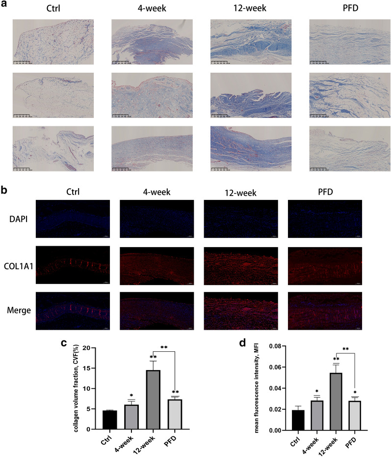
Fig. 4.
PFD attenuated the severity of synovial fibrosis (P < 0.01). Masson’s trichrome staining (a: scale bar-500 μm) and immunofluorescence staining (b: scale bar-200 μm) demonstrated that collagen deposition increased significantly in the 4-week and 12-week groups, while collagen deposition was lower in the PFD group. Quantification of collagen volume fraction (c) and mean fluorescence intensity (d) established that PFD reduced fibrosis in the synovium. Data represented means ± SD. *P < 0.05, **P < 0.01, ***P < 0.001 (the 4-week and 12-week groups were compared with the control group; the PFD group was compared with the control and 12-week groups) (unpaired t-test)

