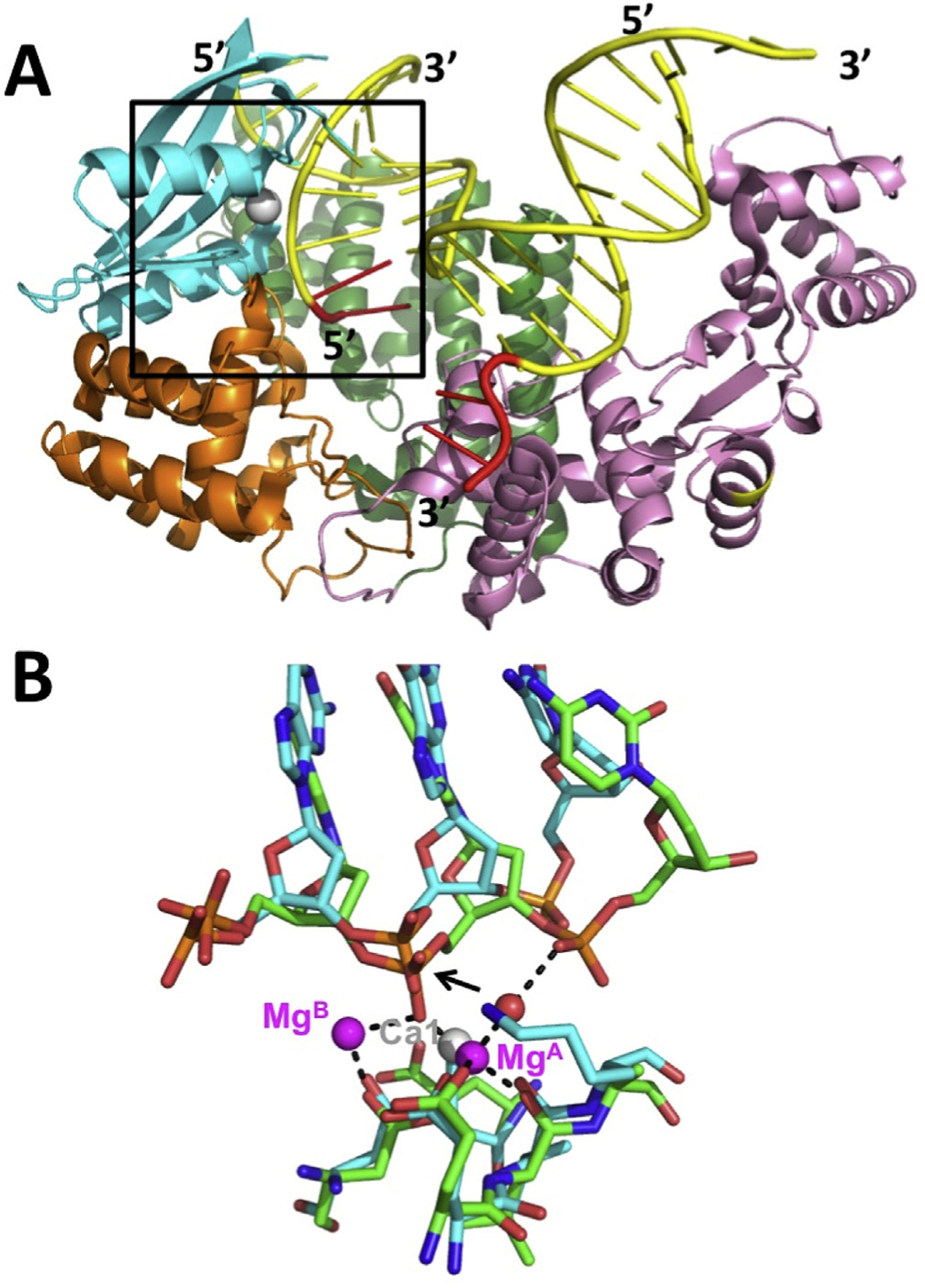
Fig. 5.

Structure of FAN1 nuclease and comparison to λ exo. (A) FAN1 structure bound to a nicked DNA (PDB code 4RI8; Wang et al., 2014). The DNA is shown in yellow, with 50- and 30-flaps in red. The T2RE domain is in cyan, with a single Ca2+ ion in grey, poised to cleave between the 3rd and 4th nucleotides from end of the 5′-flap. (B) Alignment of the active site of FAN1 (cyan bonds) with λ exo (green bonds). The 5′-3′ direction of both DNAs goes from left to right. The black dashed lines show the interactions of the metals in the λ exo structure. The single Ca2+ ion in the FAN1 structure (grey sphere) overlaps with MgA from the λ exo structure. The red sphere shows the position of the attacking water molecule, as indicated by the black arrow.
