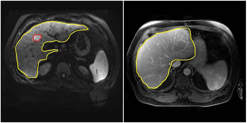
Figure 2-1:
Example images at different time points retrieved from the enterprise picture archiving and communication system (PACS) at Houston Methodist Hospital with the tumor detection by deploying U-net model [33].
(a) Automatic Liver and Tumor detection on MRI ( three months before liver transplant)
(b) Automatic Liver detection without tumor detected on MRI ( three months after liver transplant)

